|
Flea Pictures - What Do Fleas Look Like ?
you can leave one by clicking the 'donate' button opposite. The button directs you to a secure PayPal donations page where you can pay by credit card or using your own PayPal account. All tips are very much appreciated and help to fund this website. 1) What do fleas look like? - a description of the adult flea, egg, larva and cocoon (pupa). Like most other insects, fleas have three main body parts: a head, a thorax and an abdomen and, like most other insects, fleas have six legs (three on each side) that originate from the mid-section of the body (thorax). Unlike many other insects, fleas have no wings and do not fly. Perhaps to compensate for this inability to fly, the back legs of the flea are very highly developed (long and strong) compared to their other four legs and are designed for jumping. Fleas move from host to host and from host to environment to host by jumping through the air. The flea's back legs are also designed to propel the flea forwards through the animal's fur at high speed, which helps the insect parasite to evade the teeth and claws of the animal host as it attempts to remove the flea through biting, chewing and scratching activities. With the exception of the stick fast or stick tight flea, Echidnophaga (section 5), which establishes a blood-feeding position on the host (often on thinly furred or feathered regions like the legs, belly, neck, eyelids and ears) and remains there, most flea species are very mobile and are usually only seen by pet owners as brief glimpses of something brown scurrying slickly through the animal's hairs. Flea eggs are shiny, white, ovoid eggs, about 0.5mm in length. They hatch to produce grub-like flea larvae that are about 2.5mm long (depending on species). These larval fleas look like tiny white caterpillars (they move like grubs or caterpillars do too) with a black centre or core that is located acentrally, towards the head-end of the larva. Under the microscope, this black core is actually bright red and constitutes the larval stomach, which is full of digesting blood from its diet. Flea pupae or cocoons are rarely seen by owners because they are located in the pet's bedding or elsewhere in the environment (e.g. pupal fleas in the carpet), not on the pet itself. The silky cocoons are sticky and rapidly become covered in dust granules and dirt debris from the environment, making them virtually unrecognisable to owners. They just look like small balls of dirt. 2) Flea infestation pictures - a heavy infestation of cat fleas on a cat. 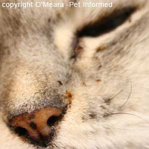 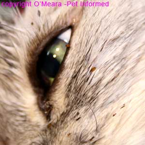
Flea pictures 1 and 2: The first cat flea picture is an image of several large fleas (Ctenocephalides felis species - the common cat flea) feeding on the face of a cat with a very heavy flea infestation. The photo's focus is on the two fat fleas located next to the cat's left nostril, however, other fleas can also be seen hanging around the eyes and forehead of the cat. The second of the flea pictures is of the eye of the same cat - fleas are located on the fur underneath the eye and one flea is sitting on the eyelid edge. These fleas are bloated and well-fed and their full abdomens appear pale brown to yellow in colour. Author's note: It is quite rare to see fleas hanging around the face of an animal unless they are stickfast fleas (these like the nose, eyelids and ears), however, the flea infestation was so severe on this cat that the fleas were absolutely everywhere. Normally, fleas prefer to crawl around the parts of the animal's skin that are difficult for the chewing, scratching host animal to reach (it makes sense - even fleas don't like to die). They therefore tend to favor the skin on the animal's inner and outer thighs, buttocks, rump, tail-base, tail, belly and back line. Author's note: This cat nearly died as a result of the severe anaemia brought on by so many feeding fleas. If you look very closely at the cat's nose (flea pictures - 1), you'll see that the nose pad is very pale brown in colour. It seems an ok colour at first glance, however, this fawn colour is just the cat's skin pigmentation, which is actually artificially masking the paleness of the nose. Look closely - there is no pink in this nose (the less pigmented parts of the nose should have a lovely rich pink shade). The cat is so anaemic and pale that there are not enough red blood cells around to make the nose appear pink. Severe infestations of fleas on kittens can be enough to kill young kittens. 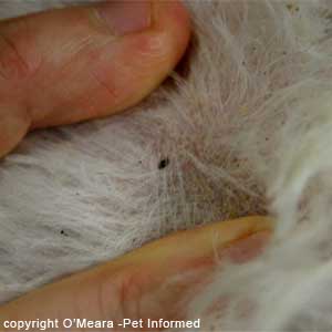 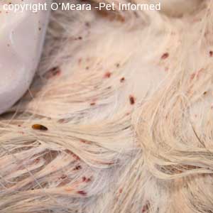 Dog fleas picture 3: This is not an image of a flea infested cat. It is a photo taken of the coat of the dog discussed in the last section of this webpage (the flea allergy dog flea pictures - section 8). The photograph contains a nice image of an adult flea crawling through the dog's fur. The fur around the flea has tiny black dots or specks that look like small bits of black dirt in the fur. This is not true dirt: these are flea droppings (flea excreta). The 'dirt' is what the flea poos out after it has digested the blood of the host animal. These flea droppings form the basic diet of the larval fleas that emerge from the flea eggs. Cat fleas picture 4: This is an image taken from the heavily infested cat pictured above. The cat's fur is wet, having just been sprayed in Frontline. A large adult flea can be seen on the left of screen. You'll notice that the fur around the flea is not covered in tiny black dots, like the dog, but in bright red smears. This is still flea excreta (what we also call flea dirt). When flea droppings get wet, the red pigment of the blood they have recently digested washes out of them, staining them and the surrounding fur red. This red is digested blood. 3) Diagram pictures of fleas - an anatomy drawing of a common adult cat or dog flea (Ctenocephalides species). 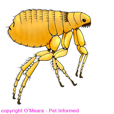 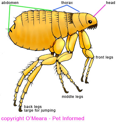
Flea pictures 5 and 6: These flea pictures contain a hand-drawn diagram of a Ctenocephalides species flea (the common house flea found on dogs and cats) showing you the basic structures of the flea's body and legs. The body has three sections: head, thorax and abdomen (labeled) and there are six legs (also labeled) that come off the thoracic section. There are no wings. I have included this diagram image on this veterinary advice flea pictures page because fleas in photographs (e.g. the microscope photos in the next section) often have legs and body parts that are greatly overlapping and difficult to discern. This overlap can make the limbs and body structures difficult to identify clearly on flea pictures. Hence the drawing - I hope it helps. 4) Flea pictures through the microscope - what do fleas look like under the microscope? 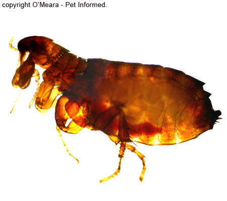 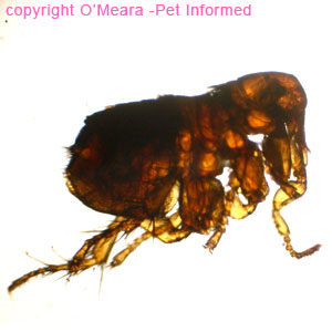
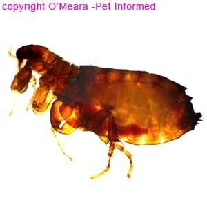
Flea picture 7: This is a microscope image of Ctenocephalides, the common dog or cat flea. It is one of the larger and more common flea species afflicting domestic pets. It will also bite humans. To give you an idea of the relatively large size of this flea, this particular image (unlike all of the other entire flea pictures presented on this page) was unable to be taken all at once, even through a low powered microscope. To provide you with a whole image of Ctenocephalides, I had to take photos of the flea in sections and Photoshop these flea pictures together to create the whole animal. Flea pictures 8: This cute, furry little flea, which looks as though it has been photoed in the very act of leaping, is the rabbit flea Spilopsyllus cuniculi. It is typically an invader of rabbit hosts, but will also cross to other host species, including the cat. This flea was taken from a cat whose hobby was going into rabbit burrows to hunt rabbits. The entire flea was able to be photographed in one shot (no Photoshop here), which suggests that this species is much smaller than Ctenocephalides. Note that Spilopsyllus cuniculi in real life might not be as dramatically smaller than Ctenocephalides as these flea pictures would suggest. The first image (Ctenocephalides) was taken of a live flea, whereas the second flea (Spilopsyllus cuniculi) was photographed after having been killed with Revolution. This death might affect its photographic size. THE HEAD OF THE FLEA - diagnosing the flea species: The head of the flea is small and simple with two eyes; a pair of basic, short, three-segmented antennae and simple, tube-like mouth-parts designed for sucking the blood of the host animal. The structure, shape and features adorning the head are important in the diagnosis of the flea species. In particular, the presence or absence of combs and the size and shape of the sucking mouth-parts can be very useful in distinguishing one flea type from another. Combs: Some species of flea (Ctenocephalides, Spilopsyllus cuniculi - flea pictures 9, 10 and 12, 13 below - and Cediopsylla simplex: an American rabbit flea - not pictured on this page) have a pair of hair-like, spiny structures called combs (or ctenidium) that are located on both the front of the face (called genal combs, these front combs look like a moustache) and on the upper thorax, just behind the back of the head (called pronotal combs, these rear combs look like the remaining hairs in a very receded hairline). As will be illustrated below, the orientation of the genal comb has a further species-diagnostic role in distinguishing Ctenocephalides from Spilopsyllus. In contrast to this, some species of flea (Echidnophaga - the stickfast flea; Pulex irritans - the human head flea and Xenopsylla cheopis - the rat plague flea) have no combs at all. The bare, combless head of Echidnophaga is seen in image 14 of the flea pictures, below. There are also species of fleas that only have a single pronotal comb and no genal comb as their facial features distinguishing them from other flea species. Mouth-parts: The stick-fast fleas (Echidnophaga gallinacea) have very long, thick, straw-like mouth-parts. These pierce deeply into the host's skin during feeding, making these fleas very difficult for the host to dislodge through biting and scratching activities. Mobile flea species like Ctenocephalides tend to have shorter, softer, weaker mouth parts that are not designed for the dual, heavy-duty roles of sucking blood and hanging on. These mouthparts just pierce the skin and suck blood when they can. They are able to be swiftly removed from the skin (so that the flea can make a run for it) when the host starts to protest at the itchy biting. 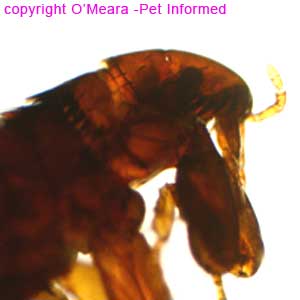 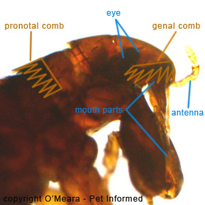 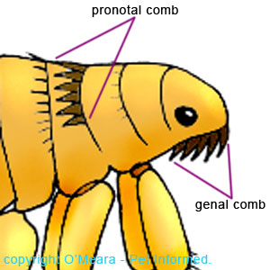 Pictures of fleas 9 and 10: These flea pictures contain a microscope photo image of the head of the adult Ctenocephalides flea (the common dog or cat flea). The simple eye (the circular brown disc in the mid-side of the face); three-segmented antennae and sucking mouth parts are marked in blue. The pronotal and genal combs are marked in brown and outlined. The presence of both combs is important if a flea is to be diagnosed as Ctenocephalides species. The orientation of the genal comb is also important in this diagnosis - the base of the genal comb of Ctenocephalides runs parallel to the flea's head and interfaces almost perpendicularly with the base of the pronotal comb. The base of the genal comb of Spilopsyllus, however, runs almost parallel with the base of the pronotal comb and at a good angle to the orientation of the flea's head. Drawing 11: This is a diagram of the head of Ctenocephalides showing the two combs. The sucking mouth-parts of the flea have been omitted from this drawing to make the combs stand out clearer.  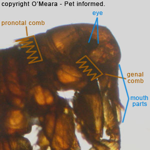
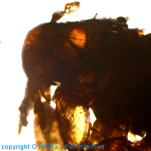
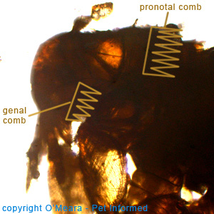
Flea pictures 12 and 13: These flea pictures contain a microscope photograph image of the head of the adult Spilopsyllus flea (the rabbit flea). The simple eye (the circular brown disc in the mid-side of the face) and sucking mouth parts are marked in blue. The antenna is there, but it is folded down next to the mouth parts so it is difficult to see (that's why it is not labeled). The pronotal and genal combs are marked in brown and outlined. The presence of both combs is important if a flea is to be diagnosed as Spilopsyllus (or Cediopsylla - another closely-related rabbit flea) species. The orientation of the genal comb is also important in this diagnosis - the base of the genal comb of Spilopsyllus (and Cediopsylla) runs almost parallel with the base of the pronotal comb and at a good angle to the orientation of the flea's head. The base of the genal comb of Ctenocephalides, however, runs parallel to the flea's head and interfaces almost perpendicularly with the base of the pronotal comb. 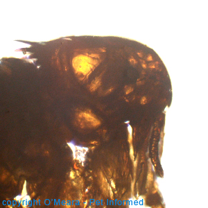 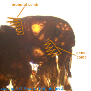
The four flea pictures above: More flea pictures of Spilopsyllus showing the unusual, characteristic genal flea comb orientation. 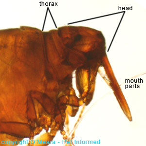
Flea image 14: This is a picture of the head of Echidnophaga gallinacea, the stick-fast or stick-tight flea of poultry and chickens. Primarily a bird flea, it will also attach to dogs, cats, people and other animals. This specimen was one of hundreds attached to an Australian echidna. The flea is distinguished by its complete lack of combs (it has no genal or pronotal combs); its long, squarish head and by its enormous sucking mouth-parts. It also has an incredibly short thorax (marked in black) compared to other more mobile flea species (compare it with the thorax images of Ctenocephalides below - flea pictures 15 and 16) and this feature makes its legs appear much more bunched together. THE THORAX OF THE FLEA: The thorax is the centre section of an insect's body. It is where the legs arise from. The shape and length of the thorax can be useful in distinguishing various species of fleas from one another. Most mobile flea species have a longish thorax, whereas Echidnophaga (image 14, above) has a very short thorax and bunched-up limbs. In some species (e.g. Xenopsylla cheopis - the rat flea) there is a telltale rod-like anatomical structure called a meral rod that is located in the mid-lower thorax just above the middle pair of legs. It is important for differentiation of this flea species. 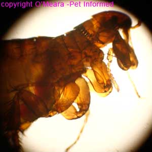 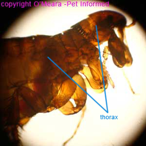
Pictures of fleas 15 and 16: These images are microscope photographs of the thorax of the common cat or dog flea, Ctenocephalides. There are no wings. THE ABDOMEN OF THE FLEA: The abdomen is the hind (rear-most) section of an insect's body. It is generally the largest section of the insect's body (i.e. it is bigger than the head or thorax parts). It contains the insect's reproductive and digestive organs as well as the series of air-filled tubules that make up the insect's respiratory system. Eggs grow and mature in the female flea's abdomen before they are laid in the animal's fur. 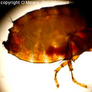 Flea pictures 17: This flea pic is a photograph of the abdomen of Ctenocephalides, taken through a microscope. The abdomen is simple with small hairs growing from it. There are no wings. THE JUMPING REAR LEGS OF THE FLEA: No picture of a flea would be complete without a photo of the flea's strong back legs. Fleas move from host to host and from host to environment to host by jumping through the air. It takes strong legs to hop like this. The flea is said to have the biggest jump of any animal, compared to its own body size! The flea's back legs are also designed to propel the flea forwards through the animal's fur at high speed so that it can evade the teeth and claws of the animal host as it attempts to remove the flea parasite through biting, chewing and scratching activities. 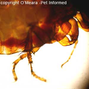 Flea photo 18: This flea picture is a photograph of the back legs of Ctenocephalides, taken through a microscope. The legs are long and strong and quite hairy. Flea pictures 19: This is a photograph of Spilopsyllus cuniculi, the European rabbit flea, mid-leap. Its hairy hind legs are so long that they stick out past the end of its body. 5) Echidnophaga gallinacea - stickfast or sticktight flea pictures. Echidnophaga gallinacea is the "stickfast" or "sticktight" flea commonly found infesting a range of poultry and chicken species. Primarily a bird flea, Echidnophaga does not seem to be too choosy about its hosts and will happily attach to the thinly furred skin regions of dogs, cats, people, horses and a range of other animals. Pet owners often find these fleas (describing them as little black or brown "dots" or tiny brown insects) clustered on the eyelids, bellies, legs, feet and ears (especially the rims or edges of the ears) of their canine and feline pets. Owners of poultry tend to find hundreds of them on the faces and necks of their birds. This particular specimen (flea pictures 20 - 22) was one of hundreds attached to an Australian echidna. The flea hangs on tightly to its host, avian or otherwise, inflicting bites that are painful and itchy and liable to becoming infected (especially in heavy sticktight flea infestations). Owners of pets often report that it is difficult to pick these fleas off the host animal, they hang onto the skin that tightly. Some people even mistakenly think that they must be ticks because they hold on to the skin so well. This ability to hang on often persists even after the flea has died (e.g. after being exposed to flea control treatments) and dead stickfast fleas often need to be scraped from the skin of the host animal to remove them. The stickfast flea is distinguished by its complete lack of combs (it has no genal or pronotal combs); its long, squarish head and by its enormous sucking mouth-parts (see flea pictures below). It also has an incredibly short thorax (marked in black on picture 21) compared to other more mobile flea species (compare it with the longer thorax images of Ctenocephalides - flea pictures 15 and 16) and this feature makes its legs appear much more bunched together. Microscope pictures of some Echidnophaga gallinacea fleas, taken from an echidna, are shown below. 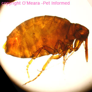
Flea picture 20: This is an image of Echidnophaga gallinacea, the stickfast flea, taken through a microscope. Notice the flea's enormous body and tiny thorax, as well as its big square head and huge, sucking mouthparts. Pictures of fleas 21: This is the head of Echidnophaga, the sticktight flea. Its head is big and square and it has enormous, piercing mouth parts. 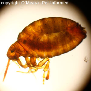
Flea image 22: This is another photo of Echidnophaga, taken through the microscope. 6) Flea larvae - pictures of larval fleas under the microscope. Flea eggs hatch to produce tiny, grub-like flea larvae that are about 2.5mm long (depending on the flea species). These newly-hatched larval fleas (called first stage flea larvae) undergo 2 molts (shed their skins) to produce second stage and third stage flea larvae (also grubs, which look very similar in appearance to the first stage larval fleas), before undergoing pupation (cocooning) to become adult fleas. Flea larvae are not that commonly seen, often falling off the host animal soon after hatching. Their part of the flea life cycle tends to occur in the pet's environment (e.g. in the carpet of the house or in the pet's bedding). These first, second and third stage larval fleas look like tiny white caterpillars (they move like grubs or caterpillars do too) with a black centre or core that is located acentrally, more towards the head of the larva. Under the microscope (larval flea pictures below: 23-25), this black core is actually bright red and constitutes the larval stomach, which is full of digesting blood from its diet of adult-flea dirt. 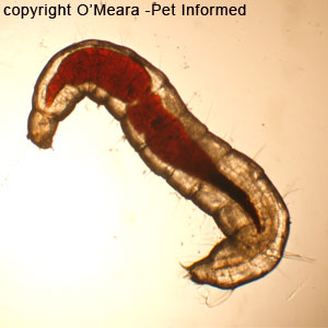 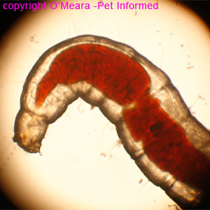
Flea larvae pictures 23 and 24: This is a larval Ctenocephalides flea as it is when seen through the microscope. The grub-like shape of the larva is clearly appreciated in image 23. The body of the grub is pale (basically white and almost see-through) with fine bristles or hairs poking out from it at intervals. The stomach of the flea larva is large and red and located towards the head of the flea larva. It is red and full of blood because, in part, the flea larva subsists on a diet of adult flea droppings (flea dirt), which are full of fully and partially digested host blood products. It is important to note that the flea larvae are not parasitic themselves, despite what these larval flea pictures might suggest. They do not feed directly from the blood of the host animal. They get their blood from the feces of the adult fleas co-existing in their surroundings. 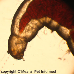
What do fleas look like - picture 25: This is the head of the flea larva. Its tiny mouth parts are visible, sticking out of the bottom of the head. A faint passage (an esophagus?) can also be imagined, running between the mouthparts and the cranial-most tip of the larval flea's stomach. 7) How can I tell if my pet has fleas - flea excreta (black specks of flea dirt) on the cat or dog's coat - evidence of flea infestation. Because fleas are so sneaky, fast-moving and photophobic (they usually avoid the light where they can and tend to hide out in denser, thicker fur) and are often present on a pet's skin only in very low numbers, many owners of domestic animals do not realise that their animal even has fleas because they do not ever see any adult fleas running around. As a veterinarian, pet owners quite often say to me, "My animal doesn't have any fleas ..." when their pet's fur is chock full of the evidence of flea activity. This oversight is completely understandable - it often takes a lot of careful, systematic coat searching in order for adult fleas to be found scurrying through the coat and, even then, this flea sighting is often very brief (the adult flea quickly slithers into another section of coat and vanishes). It is no wonder that many owners rarely ever find adult fleas on their pets, except in very high flea-burden situations. The good thing about fleas, however, is that, even though they themselves are often very skilled at keeping invisible to pet owners, they do leave behind a telltale card notifying owners of their existence: flea droppings. Every animal has to poop and fleas are no exception. The fur of a flea-infested animal will often contain tiny black dots or specks (usually around the base of the hairs) that look like small bits of black dirt or grit in the fur. This is not true dirt: these black dots are flea droppings (flea excreta). We nickname it "flea dirt", but it is not actually dirt or soil, it is tiny flea faeces. It is what the flea poos out when it has digested the blood of the host animal. These flea droppings also form the basic diet of the larval fleas that emerge from the flea eggs. Author's note: Owners often ask me how it is that I can tell whether the black dirt seen on a pet's coat is flea dirt or just plain old soil from the environment. This is easy. Firstly: look at the colour of the dirt in the fur and think about your pet's environment. Does your pet live in an environment where there is lots of fine black sand that could get into the coat? A pet that lives indoors or on good grass or on red or white sand probably shouldn't get black dirt or dust in the fur. Second: take a sample of the "dirt", put it onto some absorbable paper (a tissue is fine) and wet it and leave it to sit for a couple of minutes. If the "dirt" is actually flea droppings and thus full of blood, a red halo or corona should appear on the paper immediately surrounding and adjacent to the dirt as the blood leeches out of the droppings. Picture 28 below provides a good example of what happens to flea droppings when they become wet. 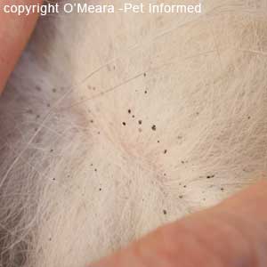 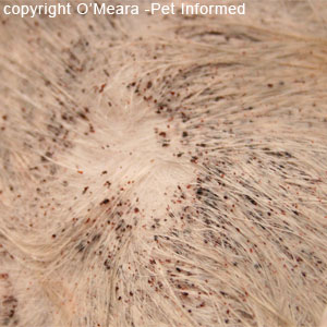
Dog flea pictures 26: This is a photo taken of the coat of a dog with a very minor flea burden. The fur of this dog contains tiny black dots or specks that look like small bits of black dirt in the fur. This is not real dirt: this is flea droppings (flea excreta). The droppings are generally situated at the base of the host's hairs because the flea moves across the skin at the base of the hairs and tends to poop where it runs. This is the flea dirt image that most owners would expect to see in a very mildly infested dog or cat. Thankfully, this dog had white fur (easy to photograph) - owners of darker furred animals will need to look very closely to see this dirt in their pet's coat. Brushing the darker-haired pet with a fine-toothed comb and examining the brushed-out particles on a white paper background can be a useful way of spotting small amounts of flea excreta. Cat flea pictures 27: This is an image taken from the heavily infested cat pictured in section 1 of this web-page. The flea dirt is extreme and shows up very well on this white cat. Not that you would have needed the flea dirt to diagnose fleas on this animal - the fleas were running up our arms as we were handling and treating the cat.
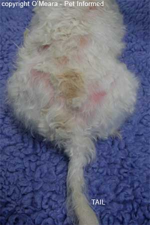
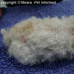
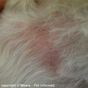
What do fleas look like? - flea dirt picture 28: This is one of the flea pictures taken from the heavily infested cat pictured in section 1 of this web-page. The flea dirt is extreme and shows up very well on this white cat. The cat's fur is wet, having just been sprayed in Frontline. A large adult flea can be seen on the left of screen. You'll notice that the fur around the flea is now not covered in tiny black dots, like the image above (picture 27), but in bright red smears. This is still flea excreta (what we also call flea dirt). When flea droppings become wet, the red pigment of the blood that the adult fleas have recently digested washes out of them, staining them and the surrounding fur red. This red is digested blood. 8) Flea allergy dermatitis and other telltale flea symptoms in cats and dogs - cats and dogs biting their bottoms red raw and over-grooming in their search of fleas. Another good way of determining whether or not an animal could have a flea infestation is to observe the animal's grooming behavior and to examine the animal's skin/fur, droppings and vomit for signs of flea-related disease or overgrooming. Grooming patterns (e.g. whereabouts on the body the animal focuses and intensifies its grooming activities); amount of grooming (time spent grooming in a day); furballs in the vomit or droppings; aggression in response to certain body-regions of handling by owners; the presence of flea-borne parasites and the presence of flea-associated regions of skin inflammation on the pet's body (see images below) can all be important clues to flea parasitism. To go from this page to the Pet Informed Home Page, click here. References and Suggested Readings: 1) Arthropods. In Bowman DD, Lynn RC, Eberhard ML editors: Parasitology for Veterinarians, USA, 2003, Elsevier Science.
Revolution is a registered trademark of Pfizer Animal Health. |
 This flea pictures page is designed to give pet owners a visual guide to common cat and dog fleas and answer the commonly-asked question: "what do fleas look like?" Everyone knows that they need to protect their cats and dogs against flea infestation, but not everyone knows what dog and cat fleas or flea infestation actually looks like. This flea pictures page contains many different photographic and diagrammatic pictures of fleas, including: images of real flea infestation, labeled diagrams of flea anatomy, microscope images of adult fleas and flea larvae, pictures of "flea dirt" (flea excreta - evidence of fleas) and pictures of flea allergy dermatitis in the dog.