Veterinary Advice Online: Cryptorchidism (Undescended Testicles).
Cryptorchidism, otherwise known as undescended testicles, undescended testes, retained testicles or undropped testes, is the condition whereby one or both of a male animal's testicles fail to fully descend into the scrotal sac (scrotum) after birth. Apart from the obvious cosmetic implications (e.g. one testicle is not acceptable in the show ring), cryptorchidism also has implications on the health, behaviour, fertility and breeding value of the affected animal. This page contains detailed information about cryptorchidism (undescended testicles) in cats and dogs (mention is also made of other animal species where appropriate).
Cryptorchidism Topics:
1. The normal process and timing of testicular descent in young puppies and kittens.
1a) The process of testicular development and descent into the scrotum in the embryonic and neonatal animal.
1b) The timing of testicular descent - when do the testicles normally drop?
1c) Why do the testes need to enter the scrotum pouch to function normally?
1d) Are some species of animals naturally cryptorchid?
2. What is cryptorchidism? - definitions and meanings.
3. How old does an undescended male animal have to be before you know its testicles will not descend and that it is a true cryptorchid?
4. How do you diagnose an animal as being cryptorchid?
4a) Diagnosing a unilaterally cryptorchid animal.
4b) Diagnosing a bilaterally cryptorchid animal.
5. What are the medical implications and complications of undescended testes?
5a) Testicular torsion (twisted testicle).
5b) Testicular cancer (testicle cancer).
5c) Male feminizing syndrome.
5d) Bone marrow hypoplasia and pancytopenia - estrogen toxicity.
5e) Excessive testosterone production.
6. What are the behavioural implications of undescended testes?
7. What are some of the economic and showing implications of cryptorchidism?
8. Should I buy (purchase) an animal if it is cryptorchid?
9. Desexing (neutering) a cryptorchid male:
9a) Why should you get a cryptorchid pet neutered?
9b) How do vets desex (castrate) a cryptorchid animal?
9c) Can the vet just remove the internal, undescended testicle and leave the descended testicle intact?
9d) Are there drugs or medications or surgical procedures available to make the retained testicle descend (i.e. cryptorchidism repair) and what are the ethical implications of this?
10. Breeding from a cryptorchid animal:
10a) Are cryptorchid males fertile and able to breed?
10b) Do cryptorchid animals exhibit typical male behaviour and libido?
10c) Is cryptorchidism hereditary? What is the mode of inheritance?
10d) Should I breed from a cryptorchid animal at all?
10e) Should I breed from the parents of a cryptorchid animal?
10f) Should I breed from the siblings of a cryptorchid animal?
10g) Is cryptorchidism more common in certain breeds?
10h) Are there drugs or surgical procedures available to make the retained testicle descend (i.e. cryptorchidism repair) and what are the ethical implications of this?
11. Is it possible to place prosthetic testicles (testicle implants) in a cryptorchid animal?
WARNING - IN THE INTERESTS OF PROVIDING YOU WITH COMPLETE AND DETAILED INFORMATION, THIS SITE DOES CONTAIN MEDICAL AND SURGICAL IMAGES THAT MAY DISTURB SOME READERS.

1. The normal process and timing of testicular descent in young puppies and kittens.
1a) The process of testicular development and descent into the scrotum in the embryonic and neonate animal.
Understanding the normal developmental processes by which each testicle forms in early embryonic life and then descends into the scrotum during late foetal life or early neonatal life (soon after birth) is very helpful to your understanding of how and why a cryptorchid state occurs. Cryptorchidism is not caused by any failure of testicular formation (the first part of this section), however the condition is a result of failure of normal testicular descent into the scrotum. Both processes will be discussed in this section.
Testicular formation and scrotal descent is a fascinating, but complicated process that I will attempt to simplify to its basics for those of you without a medical or veterinary understanding of embryology. I have made heavy use of diagrams and pictures to help you to understand.
Testicle formation and development in the abdominal cavity of the embryo:
This first image contains a simplified diagram of the internal organ structures of the developing embryo. This diagram will be our basic template for most of this section:
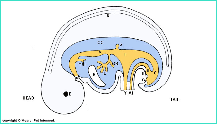
HEAD: head of embryo; TAIL: tail and pelvic region of embryo; N: neural tube (later becomes the spine and nervous system); E: eye; CC: coelomic cavity (a space that becomes the abdominal and thoracic cavities); M: mouth region; TBL: an outgrowth from the main body tube that becomes the trachea, bronchi and lungs; H: heart; S: region of the body tube that later becomes the stomach; GB: an outgrowth from the main body tube that later becomes the gall bladder; L: an outgrowth from the main body tube that later becomes the liver; P: an outgrowth from the main body tube that later becomes the pancreas; I: intestines; C: colon and rectum; B: region where bladder will form; Y: yolk sac opening; Al: allantoic cavity opening; U: region where the urethra will eventually open out (currently sealed closed); A: region where the anus will eventually open out (currently sealed closed). (Hand-drawn image interpreted and extrapolated from Dyce, Sack, Wensing - references 5, 6, 7).
The image above contains a simple diagram of an early, developing embryo in its entirety (head to tail). I have included this picture to help you to orientate yourself during this discussion because the internal structures of the embryo do not look like those of a mature animal.
Basically, the early, developing embryo has a head, a body and a tail. It has a body cavity: a space inside it (colored in blue and marked with CC - which stands for coelomic cavity, its proper name) that will later become the chest cavity and abdominal cavity spaces of the mature animal. Hanging from the roof of this body cavity by fine membranes called mesenteries is a tubelike, rudimentary intestinal-like structure (marked in orange) that will later become the bladder, intestines, stomach, liver, pancreas, oesophagus, lungs and trachea of the mature animal. This tube structure is continuous with the world outside of the embryo in three places. The first, marked M, will become the mouth of the animal. The second opening, marked Y, is continuous with the fetal yolk sac: the first source of nutrition for the early embryo prior to the maturation of the placenta and umbilical cord connections that will bring nutrients to the embryo/foetus directly from the mother's own blood. This opening (marked Y), will regress during embryonic life, once the nutrients in the yolk sac are gone, and gradually cease to be an opening at all. The third opening, marked Al, opens out into the allantoic sac, a thin, bag-like structure that holds the waste products (essentially the urine and faeces) produced by the rudimentary gut and kidneys of the embryo. This opening (Al) will regress during embryonic life and gradually cease to be an opening at all.
The formation of the kidneys and testicles:
 What you probably noticed in the last diagram was that I did not make any mention of the kidneys or reproductive organs (testicles or ovaries) of the embryo. The development of these structures will be the focus of the rest of this section before we move onto the process of testicular descent.
"Why discuss kidney formation?" I hear you ask. The reason for this is that the formation of the kidneys and gonads (gonads is general term for the testicles or ovaries) is inextricably linked. During the process of kidney formation, the embryonic body actually makes three separate attempts at producing a kidney-like excretory structure. The first attempt (a minimally-efficient kidney structure called the pronephron) occurs in earliest embryonic life and is located somewhere near the animal's head. This pronephron regresses and a second, much larger, more efficient kidney structure (called the mesonephron) is formed somewhere near the animal's chest. This structure also regresses, making way for the third and final kidney structure (called the metanephron), which will develop and grow to become the actual kidney of the mature animal. The testicle is formed during the regression of the second kidney (the mesonephron). Instead of completely regressing and disappearing, the mesonephron and many of its internal duct systems become incorporated into a new structure (the very early testicle) that is growing nearby, a fusing of the two structures (mesonephron kidney and early testicle) that eventually results in the formation of a mature and functional testicle. It is this process of renal and testicular formation that I am now going to present to you by way of a series of simple diagrams.
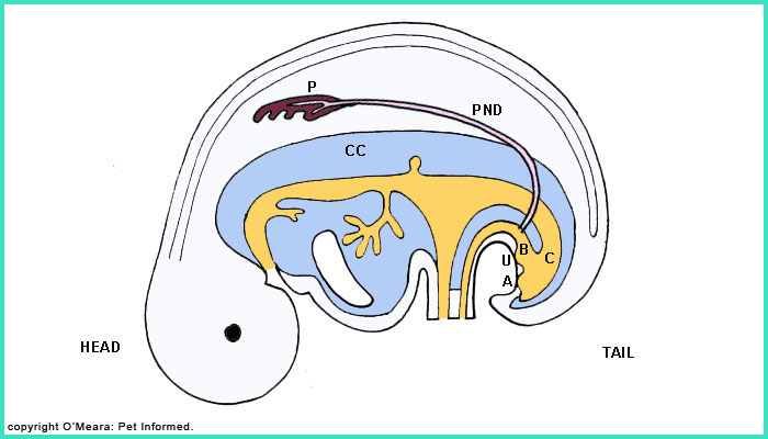
Image 1: This is a diagrammatical image of the very early embryo (same template diagram as before). The first kidneys (P: pronephron), one on each side of the body, have formed in the roof of the coelomic cavity and the watery waste products produced by their filtration of embryonic blood (i.e. urine) travel to the early bladder region (B: bladder) via tubes (one tube for each pronephric kidney) called pronephric ducts (PND).
This urine waste will be discharged out into the allantoic sac. (Hand-drawn image interpreted and extrapolated from Dyce, Sack, Wensing - references 5, 6, 7).
You will notice that the testicles have not yet made an appearance: they have not begun to form yet. You will also notice that the kidneys (as will be the testicles or ovaries) are located within the roof of the coelomic cavity, not within the cavity space itself like the other structures are. This feature persists into maturity - the kidneys and reproductive structures remain separated from the main abdominal cavity by a membranous barrier throughout life (the separate space that the kidneys and gonads occupy is termed the retroperitoneal space).
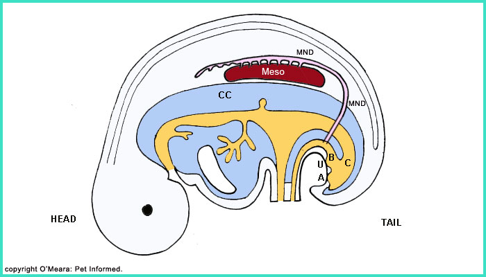
Image 2: The pronephron has regressed and the mesonephron (Meso) or second kidney has formed (one on each side of the animal). The pronephric duct (PND) has survived and has become incorporated by the filtration systems of the mesonephron to become its waste elimination conduit to the bladder region. This duct, formerly the pronephric duct, is now retermed the mesonephric duct (MND). Around the same time that the mesonephron becomes fully mature, a small bud starts growing out from the lower end of the mesonephric duct: this bud will grow to become the third and final kidney, the metanephron. We'll get to the metanephron later. (Hand-drawn image interpreted and extrapolated from Dyce, Sack, Wensing - references 5, 6, 7).
So, what about the testicles?
During the period that the mesonephron is functioning as an excretory kidney and the metanephron is just starting to bud and form, the testicles are also in the process of just starting their development. In the roof of the coelomic cavity, just medial to (closer to the midline than) the mesonephron kidneys, small thickenings of the lining of the coelomic cavity wall start to form (these thickenings will later become the thick, outer capsules of each testis). The following images explain this better.
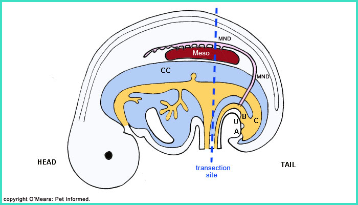
Image 3: This is the same image as image 2. You can not see the forming testicles in this view because they are being hidden from view by the large mesonephron kidney. To see the developing testicles, we will have to look at a cross-sectional view of the embryo's abdomen. The dotted line marks out where we have transected the embryonic body.
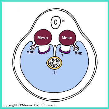
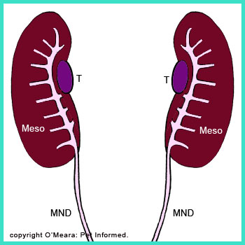
Image 4: This is a diagram of the cross-sectional view of the embryo after it has been transected along the dotted line marked in image 3. N: neural tube; Meso: mesonephron; MND: mesonephric duct; I: intestine tube and T: the early forming testicle. Also indicated is the mesentery (marked M): a thin, membranous sheet-like structure that suspends the intestinal tube-shaped structures from the roof of the coelomic (abdominal) cavity. See how closely this mesentery attaches to the region of the roof of the coelomic cavity where the new testicles are forming? This will become important (see images 8-10). (Hand-drawn image interpreted and extrapolated from Dyce, Sack, Wensing - references 5, 6, 7).
Image 5: This is a diagram of the same embryo were you to remove all of the intestinal tube and bladder structures and look up at the newly-forming kidneys and testicles from below. The mesonephron (one on each side of the animal) is the large brown structure with pink drainage tubes flowing into the pink mesonephric duct. Just medial to (closer to the midline) this mesonephron structure is the developing testicle (indicated in purple), just a thickening in the roof of the coelomic cavity at this stage. (Hand-drawn image interpreted and extrapolated from Dyce, Sack, Wensing - references 5, 6, 7).
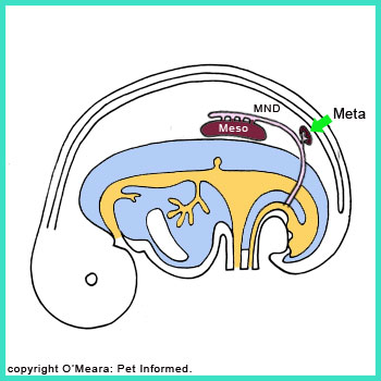

Images 6 and 7: Time passes. The mesonephron shrinks in size (starting to regress) and the third kidney (the metanephron: Meta), starts to enlarge. (Hand-drawn images interpreted and extrapolated from Dyce, Sack, Wensing - references 5, 6, 7).
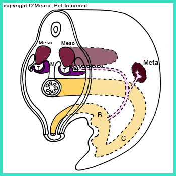
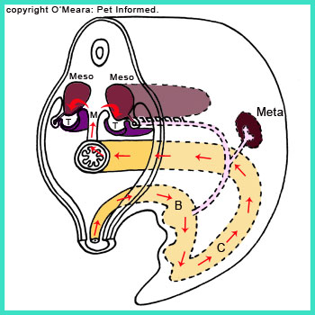
Image 8: This is a cross-sectional, oblique view of the transected embryo after it has been transected along the dotted line marked in image 3. Left and right mesonephrons (Meso) and left and right testicles are indicated as is the entire intestinal and bladder tube structure (orange) to the rear of the transection line. The mesentery (M) from which the intestinal and bladder structures hang is also indicated.
Image 9: This diagram is the same one shown in image 8. It demonstrates how there is a pathway running from the outside of the embryo (yolk sac and allantoic region) to the region of the testicles. This path is marked in red arrows. (Hand-drawn images interpreted and extrapolated from Dyce, Sack, Wensing - references 5, 6, 7).
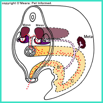
Image 10: Germ cells (kind of like stem cells) travel from the yolk sac region to the developing testicle bumps along this pathway (path indicated in images 8 and 9). These germ cells will take up residence in the developing testicles and become the spermatogenic cells that divide and mature throughout the male animal's life to produce the sperm. (Hand-drawn image interpreted and extrapolated from Dyce, Sack, Wensing - references 5, 6, 7).
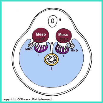
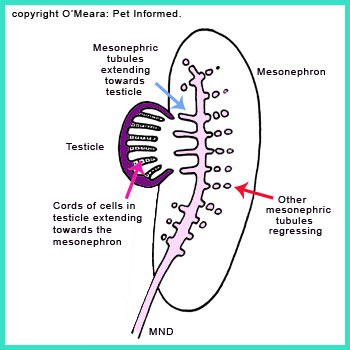
Image 11: Cords of cells grow inwards from the surface of the testicular thickenings. These cells will later mature and rearrange themselves to become the duct systems and the hormone-producing (e.g. testosterone producing), sperm-producing and sperm-maturing cell populations of the mature testicle.
Image 11 and 12: As the cords of testicular cells (indicated in image 11) are forming, some of the filtration duct tubes (called mesonephric tubules) from the now-regressing mesonephron kidney, adjacent to the testicle, also start to elongate, stretching out from the mesonephric duct (MND) towards the newly developing testicle. These filtration ducts will eventually fuse with the cords of cells developing in the testicle, becoming a series of testicular ducts that will drain into the main mesonephric duct and out into the bladder region. The other filtration ducts (mesonephric tubules) of the mesonephric kidney that are not incorporated by the testicular drainage system will regress and disappear (image 12). (Hand-drawn images interpreted and extrapolated from Dyce, Sack, Wensing - references 5, 6, 7).
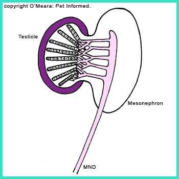
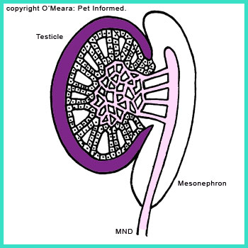
Image 13: A diagram image of the cords of testicular cells uniting with the filtration ducts (mesonephric tubules) of the original mesonephric kidney to form continuous ducts from the testicle to the mesonephric duct (MND).
Image 14: The mature testis. The cords of testicular cells have organised themselves into complex glandular structures capable of producing spermatozoa (sperm) and hormones. A complicated meshwork of ducts (called the rete testis) runs through the centre of the testicle collecting sperm before draining into the mesonephric duct (now termed the epididymus and vas deferens/deferent duct) via a series of small ductules (termed efferent ductules). (Hand-drawn images interpreted and extrapolated from Dyce, Sack, Wensing - references 5, 6, 7).
The testicle, large and heavy now, will start to hang from the roof of the abdominal cavity by membranes (mesenteries), similar to the intestine. In the female animal, the right and left ovaries and their respective duct systems will actually migrate down the walls of the abdomen, the ducts fusing in the centre of the animal's body between the colon and bladder to form the uterus. What this means is that, even though the testicles and ovaries/uterus are formed in the roof of the coelomic cavity and are essentially retroperitoneal in location, because they hang down from long mesenteries (these mesenteric membranes are essentially out-pouchings or "slings" of the dorsal coelomic cavity lining: the membrane that divides the abdominal space from the retroperitoneal space) they do tend to take up positions well within the abdominal cavity of the animal, rather than remaining tightly within and against the roof of the abdominal cavity like the kidneys do.
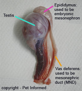
Image 15: This is an image of a canine testicle that has been removed from a dog during castration. You can still see the parts that were once the mesonephric kidney and forming testicle bulge.
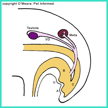
Image 16: In this picture the testicle has completely formed. The mesonephron has become incorporated into the testis and no longer functions as a kidney and the mesonephric duct (now a conduit for sperm, not urine) is now termed a vas deferens or ductus deferens or deferent duct (VD). The metanephron (Meta), the third and final kidney, now takes over the role of blood filtration and waste product excretion. (Hand-drawn image interpreted and extrapolated from Dyce, Sack, Wensing - references 5, 6, 7).
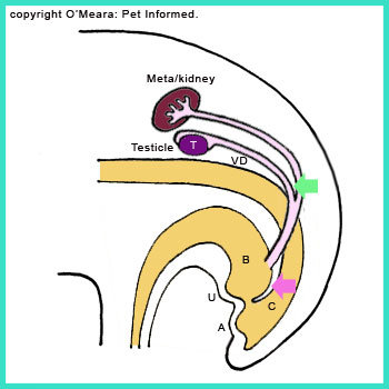
Image 17: The metanephron enlarges and moves forwards (cranially) towards the animal's chest. At the same time, the testicle, pulled by the gubernaculum (the gubernaculum will be explained in the testicular descent part of this section), starts to move toward the animal's rear. A cleft or division starts to form (green arrow) between the metanephric duct (MetaND) and the vas deferens. A cleft also starts to develop (pink arrow) between the colonic (C) and bladder (B) regions of the intestinal-bladder tube. (Hand-drawn image interpreted and extrapolated from Dyce, Sack, Wensing - references 5, 6, 7).
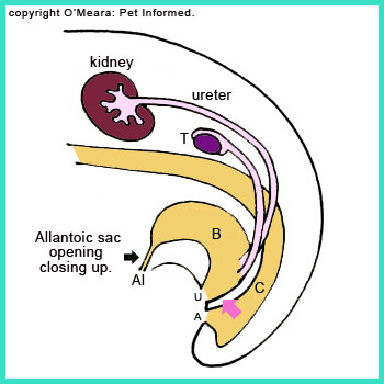
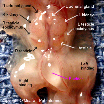
Image 18: The metanephron kidney enlarges more and moves into its final resting place. The testicle continues to be pulled caudally (rearward) by the gubernaculum. The metanephric duct (now called the ureter) divides completely from the vas deferens and will occupy a position in the neck of the developing bladder. The vas deferens will occupy a place further down, in the region of the animal's prostate and urethra and not in the bladder (urine is bad for sperm survival and so it would be bad for the male animal's fertility if the vas deferens ended up discharging into the bladder). The colon and bladder form and divide away from each other (pink arrow) to open up via different holes (the anus: A and urethra: U). The seals over the urethra and anus regions break down, allowing the fetus to urinate and defecate through these holes. With the bladder waste (urine) now able to drain out via the urethral opening (U) and the intestinal waste (feces) now able to drain out via the anal opening (A), the Allantoic sac opening (Al) is now no longer needed. It shrinks and regresses (black arrow). (Hand-drawn image interpreted and extrapolated from Dyce, Sack, Wensing - references 5, 6, 7).
Image 19: This is an image of the fully-developed reproductive and urinary structures of a male kitten fetus (2nd trimester). This picture is a close-up of the roof of the embryo's abdomen, following removal of all of the intestinal and liver structures. In this image, the kidneys are large and clearly visible in their final mature-animal positions and the formed testicles and epididymi are visible just below them. The bladder (clear, round structure) has been flipped downwards to reveal these structures. Notice how the tip of the bladder (near bottom of image) is pointed and narrow: this is the allantoic sac opening just starting to regress.
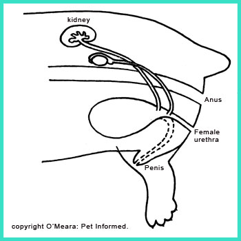
Image 20: The intestinal and urethral structures rearrange position as the foetus matures and develops limbs and bones and proper pelvic structures. The urethra, instead of opening out just under the anus as would occur in a female animal (Female), becomes incorporated into the formation of the male penis. This results in an elongated urethra that, in the dog and horse and other male animals, is positioned under the animal's belly (forms the penis). On this diagram, this penile urethra is indicated with a dotted line. Note that the formation of the male penis is not as simple as the urethral passage just elongating: it is actually a much more complicated process that will not be discussed in further detail here because it does not affect your understanding of cryptorchidism.
The process of testicle descent:
In order for the sperm produced by the animal's testicles to be fertile, the testicles must relocate to a cooler site, well outside of the animal's hot body core (i.e they must relocate to a special testicle-holding pouch just under skin behind the penis, which is called the scrotum). This process, called testicular descent, occurs much later than testicular development: testicular descent occurs in late foetal or early neonatal life (i.e. soon after birth), whereas testicular formation occurs much earlier as an integral part of early embryonic development. Even though it is only this process of testicular descent that really matters with regard to your understanding of cryptorchidism (i.e. this part of section 1a), you do need to have some understanding of how it is that the testicles start their life in the abdomen and some idea of where they come from in order to understand the testicular descent process fully. Hence the detailed description of gonad (testis) formation provided in the first part of this section. Once again, I have used simple diagrams as a way of illustrating the basic principles of testicle descent.
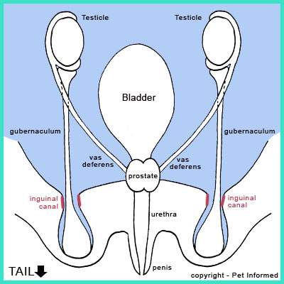
Image 1: By the time we get to the process of testicular descent, most of the fetus's organ systems will have developed to their final stages and now be recognizable as the organs that are present within the adult, fully mature animal. In the male foetus, reproductive and urinary structures such as the bladder, kidneys, ureters, urethra, penis, prostate, vas deferens and testicle will all be recognisable. The only incomplete developmental process is that the testicles are still located inside of the foetus's abdominal cavity (as seen in this image) and not inside the scrotal sac. (Hand-drawn image interpreted and extrapolated from Dyce, Sack, Wensing - references 5, 6, 7).
What has not yet been mentioned on this page is that, during the process of testicular development, a tight band of connective-tissue, ligament-like material (termed the gubernaculum) forms which links one end of each testicle to the region underneath the animal's perineal skin (skin between the anus and penis) where the corresponding (right or left) scrotal sac will be. The path of the gubernaculum band, from testis to scrotal site, passes through a hole (termed the inguinal canal) in the muscular wall of the animal's abdominal cavity (coelomic cavity), somewhere in the region of the animal's groin. The gubernaculum's role is to contract and draw the testicle from the abdominal cavity through the inguinal canal and into the scrotal sac. To be more specific, the gubernaculum draws the testicle from where it hangs within the abdominal cavity by sliding it along the retroperitoneal space (remember that the testicle is retroperitoneal in location - it hangs down from the roof of the abdominal cavity, slung within the membrane or mesentery that divides the retroperitoneal space off from the abdominal cavity space).
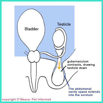
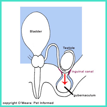
Images 2 and 3: The gubernaculum is contracting, dragging the testicle from where it hangs within the abdominal cavity, through the inguinal canal (a hole in the wall of the abdominal cavity, which is also continuous with the aforementioned retroperitoneal space) and into the scrotal sac. (Hand-drawn images interpreted and extrapolated from Dyce, Sack, Wensing - references 5, 6, 7).
During this process, some of the lining of the abdominal cavity also gets drawn (dragged) into the scrotal sac along with the migrating testicle (I have attempted to indicate this in diagrams 1-4 by coloring the abdominal space component of the scrotal sac in blue like the coelomic cavity). In image 2, you can clearly see that the scrotal space (where the testicle will sit) contains a small out-pouching of the lining of the abdominal cavity: i.e. some of the scrotal space where the testicles will sit is continuous with the animal's abdominal cavity.
Knowing that much of the scrotal pouch is essentially continuous with the animal's abdominal cavity is important for two reasons. One: it explains why a cryptorchid or undescended testicle can be discovered anywhere from the abdominal cavity to just ahead of the animal's scrotal sac, including within the inguinal canal itself, and two: it also explains why some animals develop scrotal or inguinal hernias (also called testicle hernias) - a condition whereby the intestines or other abdominal cavity contents of the animal migrate through the inguinal canal of the animal and become entrapped within the animal's scrotal sac (this results in a very painful and swollen scrotal sac and often requires urgent surgical correction).
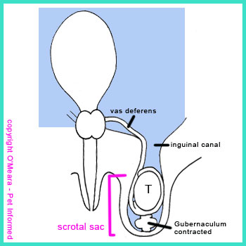
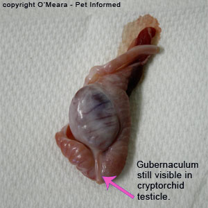
Image 4: The gubernaculum finishes its contraction, having completely drawn the testicle into the scrotal sac. Once the gubernaculum band has finished contraction, it completely regresses, shrivelling up to not much more than a small bleb of scar tissue located at the bottom pole of the testicle. Prior to complete gubernacular regression, it is possible for the testicle to slide back and forth between the abdomen and scrotal region (you often see this in young puppies - the testicles are very mobile and able to be pushed away from the scrotal sac and up towards the inguinal canal). After gubernacular regression, the testicle is fixed within the scrotal sac and is unable to move out of it, at least in those species where testicle retraction is not a desired possibility. In section 1d, I discuss animal species that are able to retract and move their testicles from their scrotal sacs into their abdominal cavity and back again. (Hand-drawn image interpreted and extrapolated from Dyce, Sack, Wensing - references 5, 6, 7).
Image 5: This is a picture of an intra-abdominal cryptorchid canine testicle that was removed at surgery. You can clearly see the fibrous tissue band extending from the tail of the testis. Prior to surgical removal, this band ran from the tail of the testis, through the inguinal canal, to the inside of the scrotal sac. This fibrous band is called the gubernaculum.
1b) The timing of testicular descent - when do the testicles normally drop?
The timing of testicular decent, for those species whose testicles do descend, differs from species to species and possibly even from breed to breed within a species.
Cats:
In feline kittens, the process of testicular descent normally occurs just prior to birth (i.e. whilst the male kitten foetus is still in its mother's womb). Therefore, tom kittens should have tiny testes palpable in their scrotums at or shortly after birth.
Dogs:
In canine puppies, the process of testicular decent normally occurs shortly after birth (within 5-10 days of birth) and most male pups will have readily palpatable testicles within their scrotal sacs at around 10-14 days of age. I have found that most male puppies have readily palpatable testicles in their scrotum sacs by the time of first vaccination (6-8 weeks).
Because the gubernaculum (see section 1a) may not be fully receded and contracted down in some 6-7 week-old pups, it is likely that these testicles will still be mobile and able to slide back and forth between the puppy's abdomen and scrotum. Puppies will often retract these mobile testicles into their abdominal cavities when they are excited or anxious (e.g. they are often excited or nervous at vaccination time). As a result, some pups aged 6-7 weeks and earlier are wrongly diagnosed as being cryptorchid (retained testicles) because their retractile testes are being sucked up (retracted) into their abdominal cavities during excitement. The gubernaculum generally recedes (shrinks) fully, preventing the movement of the testicle from the scrotum to the abdominal cavity or inguinal canal, by about 8 weeks of age and, after this point, the testes are either palpable within the scrotal sac or they are considered retained (cryptorchid) - see section 3 for more details on the timing of testicle descent with regard to the diagnosis of true cryptorchidism.
Author's note: You will occasionally get some pups who, at 8-10 weeks of age, will still be exhibiting slight testicular movement: they will be able to pull their testicles forwards, alongside the penis, when stressed or excited. These pups should be able to drop their testicles back into their scrotum when relaxed and it should not be difficult for the vet to manually manipulate the testicles back into the correct scrotal position during the examination. Testicles that are difficult to move back into place should be suspected of having cryptorchidism.
Author's note: The timing of testicular descent has not been studied in every individual breed of dog. There may be some breed variation in the timing of testicular descent. For information on the testicular descent of individual breeds, it may be best to look up a specific breed site or ask a couple of dog breeders who breed that kind of animal.
1c) Why do the testes need to enter the scrotum to function normally?
Spermatogenesis: the production of viable sperm, is greatly affected by the temperature of the testicle (the testicle is the site of sperm production) in many species, including the dog, cat, horse and most livestock animals. In many species, the aforementioned animals included, the temperature inside the animal's abdominal cavity or inguinal canal region is simply too high for the production of viable sperm to occur should the testicles remain within these body regions. In these species, the testicles need to descend to a site just under the skin and far from the hot abdominal cavity (i.e. the scrotum) in order to attain a lower and cooler temperature; one that is appropriate for successful viable sperm production.
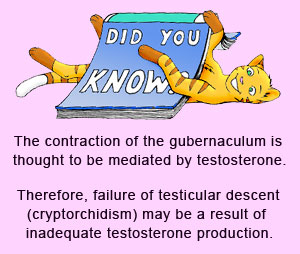
The production of testosterone (main testicular hormone) is not affected by temperature and, as a result, undescended testicles will still make plenty of this "male hormone" regardless of the fact that the undescended testicle itself is effectively infertile.
Author's note: The production of viable, fertile sperm is so sensitive to increases in temperature that sperm production can drop even in a fully descended, scrotal testicle if there is too much hair or wool covering the scrotal sac or if inflammatory processes occur in and around the testis and epididymus. Orchitis (testicle inflammation) and epididymitis (inflammation of the epididymus) both result in a hot, swollen testis (site of sperm formation) or epididymus (site of sperm maturation) respectively. This inflammatory heat can greatly reduce the output of viable sperm.
1d) Are some species naturally cryptorchid (have undescended testicles)?
For reasons unknown to us, there are some species that do maintain a naturally cryptorchid state for either all or part of the year. Animals such as birds, elephants and their closely-related cousins: the hyraxes, do not descend their testicles throughout their lives. These species do not seem to have any trouble breeding, which suggests that their spermatogenesis is able to occur regardless of internal body temperatures or that they have found other ways of cooling their testicles besides swinging them in the breeze. Many rodent and lagomorph species (rabbits, hares, mice, rats, guinea pigs, cavies) have readily mobile retractile testes that are able to move back and forth from the scrotal sac to the abdominal cavity at will. These species are readily able to retract their testicles into their abdominal cavities during veterinary examinations, a feature which can sometimes make these species difficult to sex. It is thought that some of these species, which have a distinct breeding season, may keep their retractile testicles undescended (infertile) during the off-season and only descend their testicles just prior to and during the mating season, when viable sperm production is required.
2. What is cryptorchidism? - definitions and meanings.
 Cryptorchidism is defined in most textbooks as a failure of normal testicular descent. In the dog, the definition has been further refined and canine cryptorchidism is defined as the condition whereby one or both testicles are not present within the scrotal sac of the canine animal by the age of 8 weeks. The term encompasses situations whereby the testicles are still located within the male animal's abdominal cavity and also situations whereby the testicles have exited the animal's abdomen, but are residing in the animal's inguinal ring (inguinal canal) or regions underneath the skin, lateral to the penis and just cranial to (ahead of) the scrotal sac.
Some textbook sources say that you can not 100% diagnose a pet cat or dog as being cryptorchid until it has reached puberty: 6 months for cats and smaller dog breeds and around 12 months for large breeds (theoretically, puberty occurs at around 18 months for giant breeds, but they rarely get the condition anyway). Certainly if the animal gets to 1 year of age (which should be prior to that male animal being permitted to breed anyway) and has not dropped, then it should be considered exceedingly unlikely to. In fact, most textbooks claim that if the animal has not descended by 2 months (8 weeks) of age then it is very unlikely to, however, because you do occasionally come across the rare individual animal that drops late, some of these texts do permit the puberty age cut off.
Cryptorchidism is divided into two subgroups:
Unilateral cryptorchidism - unilateral cryptorchidism is the condition whereby only one testicle is within the scrotal sac of the male animal, but the other one is retained and undescended. Unilateral cryptorchidism is easy to diagnose because you can generally be certain that the animal has not been desexed (you can feel the scrotal testicle).
Bilateral cryptorchidism - bilateral cryptorchidism is the condition whereby neither of the testicles is within the scrotal sac: both are retained and undescended. Bilateral cryptorchidism can sometimes be difficult to distinguish from a desexed male in the pre-surgery animal, particularly if the animal has had an uncertain history (i.e. it was obtained from a shelter, has not been owned by this current owner since it was a kitten or puppy, the animal has had several owners and so on) and its desexing status is unknown.
Conditions that are sometimes confused with cryptorchidism:
Cryptorchidism is different to monorchidism (also called monorchism) and anorchism, both of which are very rare in comparison to cryptorchidism.
Anorchism is the condition whereby a male animal has been born completely without testicles (i.e. it truly has no testicles). It is questionable whether such an animal that is completely missing testicles should be termed "male" in the true sense. Such an animal is likely to have major sex chromosome defects and will most likely show other significant body changes including: persistently immature sexual behavior and poor, absent or malformed development of secondary genitalia.
Monorchism is the condition whereby a male animal has been born with one testicle only (i.e. the animal just has a single testicle). The veterinarian often performs surgery, believing the animal to be a unilateral cryptorchid, but never finds a second testicle on surgical exploration.
3. How old does an animal have to be before you know that the animal is a true cryptorchid and that its testicles will not descend?
For most general pet-owners, knowing an exact age, beyond which an animal with undescended testicle/s must be considered a cryptorchid, is a moot point if their intention is to get that male dog or tomcat desexed regardless (for health reasons, people should get their male dogs and cats desexed if they have no intention of breeding).
For commercial and showing breeders of dogs and cats, however, the age definition of cryptorchidism is very important because it has a bearing on whether a young dog or tomcat will become a stud animal (breeding animal) or show animal in the future.
Most texts define cryptorchidism as the condition whereby one or both testicles are not present within the scrotal sac of the animal (dog or cat) by 8 weeks of age. Using this definition, animals whose testicles have not descended into the scrotum by this time should ideally not be bred from and should be considered cryptorchid. Certainly, many of the textbooks state that if a male animal's teste/s have not descended by 2 months of age, then they are very unlikely to. Other texts are a little more generous, allowing the animal up to 16 weeks before deeming the animal's gonads unlikely to descend. This 16 week timing is supported by the findings of those veterinarians who have trialled medical therapies to induce pet testicles to descend. Those vets who have had some success in inducing retained testicles to descend, using drugs and medicines, have found that success is sometimes achieved if the treatments are administered prior to 16 weeks of age (although it is not 100% certain if the testicles would have dropped anyway regardless of treatment given), but that medications given after 16 weeks of age usually resulted in no effect on testicular descension. For more on the medical induction of testicular descent, see section 9e.
Many breeders are loathe to cull an otherwise excellent quality (good temper, good features etc.) stud dog or tomcat from their breeding pool, just because its testicles have not descended by 8 weeks, because they have heard of cases where the testicles have come down months later. Late testicular descent certainly can occur (though it is rare). I have personally heard of a case whereby a cryptorchid show dog was given a prosthetic testicle to hide the fact that one of its testicles was not descended, only for the true testicle to descend later on, resulting in the show dog having three testicles for a brief period of time before the prosthesis was removed.
Some of the textbooks cover their bases on the matter of delayed testicular descent by claiming that you can not 100% diagnose a pet as being cryptorchid until it has reached puberty: 6 months for the cats and smaller dog breeds and around 12 months for the large breeds (theoretically, puberty occurs at around 18 months for the giant breeds, but they rarely get the condition anyway). Certainly, if the animal gets to 1 year of age (which should be prior to any male animal being permitted to breed anyway) and one or both of its testicles have not dropped, the general opinion is that they are very unlikely to and that the animal in question is, therefore, a true cryptorchid.
Those of you breeders who are unwilling to cull out stud males on the basis of the 8-week cryptorchidism definition, or the more generous 16 week definition given by some authorities, I advise to hold off on breeding your male animal until it has reached the 1 year of age mark (you probably shouldn't be mating a male dog or cat before this age anyway). If the testicles have descended by this time (1 year), then the animal may just have been a late descender and not a true cryptorchid. If the testicles have not dropped by this time (1 year mark), then it is most likely the testicles will never descend and that the animal in question is therefore a true cryptorchid.
4. How do you diagnose an animal as being cryptorchid?
As a general rule, it is very easy for veterinarians to diagnose an animal as being unilaterally or bilaterally cryptorchid if they have seen the animal since it was a young pup or kitten (i.e since first vaccination). After all, in this kind of situation, the veterinarian gets to palpate the young male's scrotum at weeks 6-8, 12 and 16 and, if the testicles have not descended by the third visit, then it is very likely that the animal can be termed a true cryptorchid (unless it ends up being one of the rare late descenders discussed in section 3). In a similar fashion, it is usually easy for the vet to diagnose an animal as being cryptorchid if it has been with the one owner for all of its life (since kitten-hood or puppy-hood), even if it has not been seeing the one vet. Most owners can usually remember if their pet has been castrated or not. The only time that this theory may not hold up is if a neighbor has taken exception to an entire tomcat or dog worrying at his females or peeing on his lawn and has smuggled the culprit off to the vet for a quick snip without the owner knowing about it (it happens).
The main difficulty in diagnosing an animal with cryptorchidism comes when the animal's history is unknown. The animal might have been a stray when obtained or have been through several different owners, any of whom might have gotten the male castrated without the current owner being made aware of it. The following sections (4a and 4b) provide information on how veterinarians and owners can diagnose an animal with unilateral or bilateral cryptorchidism respectively.
4a) Diagnosing unilateral cryptorchidism in an animal.
1. History from owner:
If the owner has had the animal since it was very young or the pet has been in the owner's family since it was very young, it is unlikely that the animal will have had a single undescended testicle removed without the owner being aware of it (particularly if the normal testis is still in place).
If the animal has had several owners in the past, it may not be possible to tell from the history given by the current owner whether or not the animal is a true unilateral cryptorchid or if it has had a previous surgery to remove the missing, presumed-undescended, testicle. Veterinarians should still ask the question about previous owners' activities, however, because the current owner may well know some of the previous owners and these people might be able to be contacted to provide a more detailed surgical history on the animal.
2. The breed:
The breed of dog or cat may well increase or decrease a veterinarian's suspicions about whether or not it could be cryptorchid. Certain breeds (particularly pure breeds) of dog and cat have a higher incidence of cryptorchidism, whereas certain other breeds have a low incidence of the condition. Dog breeds at high risk of cryptorchidism tend to be small breeds including: Toy and Miniature Poodles, Pomeranians, Chihuahuas, Malteses, Cairn and Yorkshire Terriers, Dachshunds, Schnauzers, shelties (Shetland Sheep dogs), Pekingeses and Bulldogs. Larger breeds, in particular the Boxer and Old English Sheepdog, may also be affected. In the cat world, the Persian is most commonly implicated, however, we have seen a number of domestic short hair and longhair cats (moggies) with the condition in our clinic. Large dog breeds seem to have a greatly reduced risk of cryptorchidism. Those at low risk include the Saint Bernard, Great Dane and various field and hunting dogs like the Golden Retriever, Labrador, Beagle and English Setter. Mixed breed (mongrel) dogs are thought to have a lower incidence than pure breed dogs.
3. Physical examination and testicle palpation:
Diagnosing an animal with unilateral cryptorchidism is generally very simple and can be often be done on the basis of a simple physical exam and testicle examination. The veterinarian will palpate the animal's scrotum during a simple health check or pre-anaesthetic check-up (e.g. prior to the animal being castrated) and, instead of finding two testicles in the scrotal pouch, only find just one testicle. Given that it is very rare for a previous veterinary surgeon to only remove the one testicle or to only remove a retained testicle without taking the scrotal sac testicle, the assumption, upon finding only one testicle in the bag, is that the other testicle is undescended. Thus, the animal is diagnosed as a unilateral cryptorchid.
The diagnosis of unilateral cryptorchidism is made particularly easy when the retained testis is located outside of the animal's abdominal cavity and is somewhere within the inguinal ring region or prescrotal region. In these cases, the undescended testis is often able to be palpated, particularly if the animal is lying on its back. The undescended testicle is felt as a lump under the skin on testicle exam. If the animal is under a general anaesthetic (e.g. prior to castration) it is even easier.
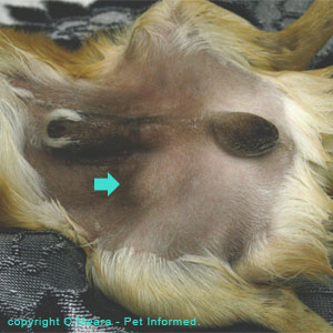
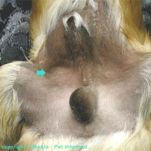
Pictures: The above pictures are photos of a Pomeranian dog with unilateral cryptorchidism. This cryptorchid dog has a large scrotum with a single testis inside. The other testicle (the dog's right testicle) has not fully descended and can be seen as a bulge just under the skin, located alongside the dog's penis and prepuce. It is marked with a green arrow.
4. Examining the animal for scars:
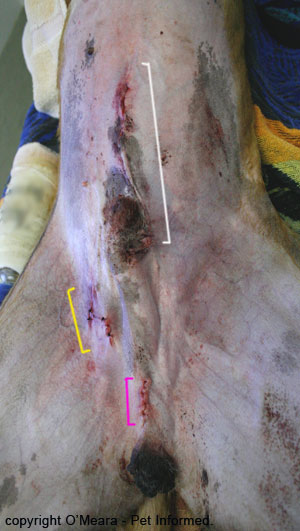
Occasionally, you will come across cases whereby a previous vet has removed a single scrotal or abdominal testicle and left the remaining scrotal testicle intact. These cases look very much like unilaterally cryptorchid dogs and cats, except that the second testicle can not be found at surgery (next point). You might get a clue that this has happened by looking for obvious past surgery scars in the regions of the prescrotum (just in front of the scrotum), inguinal region and/or midline abdomen. The image below provides a visual reference to where these scar lines might be found.
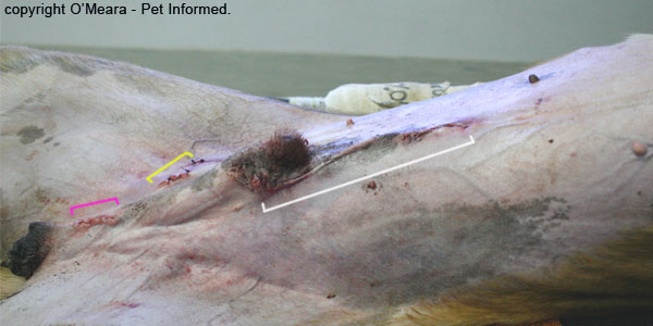
Picture: The above image is a photograph taken of a dog immediately following a surgery to locate a unilaterally-missing testicle. The penis is the small, dark, hairy mound in the centre of the image and the scrotal sac is the black lump on the left hand edge of the image. The dog's head is towards the right of this picture. The suture line indicated in pink is the site through which the scrotal testicle was removed during the castration. The suture line indicated in yellow is the incision made into the inguinal region where one might find an undescended testicle lodged in the inguinal canal. The suture line indicated in white and running alongside the front edge of the animal's penis is the incision made into the abdominal cavity where one might find an abdominally located undescended testicle. Scars in any of these areas might indicate that a prior castration or cryptorchid-castration surgery has been performed.
5. Exploratory surgery:
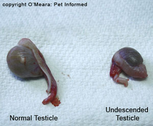 Sometimes a diagnosis of unilateral cryptorchidism (as opposed to monorchism or prior surgical removal of a single testicle) can only be made by surgical exploration. In these situations, the animal is placed under a general anaesthetic and its entire abdomen, groin and scrotal region is shaved and surgically prepared. Because the missing testicle might be very small (the retained testicle is often a lot smaller than the scrotal testicle) and could be located anywhere from the abdominal cavity, behind the kidney, through the inguinal canal to the prescrotal region, the vet may have to make several incisions to find it (all this in addition to making an incision into the skin just ahead of the scrotal sac to remove the descended testicle - pink line on the photo below). The vet might make an incision into the abdominal cavity (white line on the photograph below), only to discover that the retained testicle's spermatic cord (vas deferens and testicular blood vessels) disappears into the inguinal canal. The vet will then need to suture up the midline abdomen incision and make a new incision into the inguinal (groin) region (yellow line on the photograph below) to locate the missing testicle.
Picture: The above image is a photograph taken of a dog immediately following surgery to locate a unilaterally-missing testicle. The penis is the small, dark, hairy mound in the centre of the image and the scrotal sac is the black lump on the bottom edge of the image. The dog's head is towards the top of this picture. The suture line indicated in pink is the site through which the scrotal testicle was removed during the castration. The suture line indicated in yellow is the incision made into the inguinal region where one might find a testicle lodged in the inguinal canal. The suture line indicated in white and running alongside the front part of the animal's penis is the incision made into the abdominal cavity where one might find an abdominally located testicle.
Author's note: Occasionally a previous veterinarian, unbeknownst to the current owner, will have removed only one of the animal's testicles, leaving the other one intact. This is generally considered to be bad practice and occurs for a couple of reasons: the veterinarian removed a retained testicle to prevent it from becoming cancerous (see section 5) and left the descended testicle intact or the veterinarian removed a single scrotal testicle because it was diseased or injured or requested by an owner (e.g. Greyhound racing dog owner) and left the other one intact (making the pet appear falsely cryptorchid). In this situation, the veterinarian will diagnose the animal as being unilaterally cryptorchid on testicular palpation and testicular examination, but find absolutely no testicle (just the stump of the ligated vas deferens and testicular blood supply) at surgery.
In the case of the greyhound dog, pictured above, no retained testicle was found, only a ligated testicle stump. This dog had had its testicle removed by a previous vet. This would have been nice to know beforehand so that we might have avoided causing all of that surgical trauma.
Author's note: In the case of a truly monorchid animal (an animal born without one testicle), there will be no fully developed vas deferens or testicular blood supply to the missing testicle discovered at surgery. Both of these structures will be completely absent, along with the undeveloped testicle.
4b) Diagnosing bilateral cryptorchidism in an animal.
1. History from owner:
If the owner has had the animal since it was very young or the animal has been in the owner's family since it was very young, it is unlikely that the animal will have had its testicles removed (i.e. been neutered or castrated) without the owner being aware of it. The only time that this may occur is if a neighbour takes exception to the male animal (dog or tom cat) roaming, urinating on his flowers or worrying at his bitches or queens. Such a neighbour might well smuggle the culprit male off to the vet for a quick snip without the vet being made aware of the fact that the cat or dog in question is not the property of that owner.
If the animal has had several owners in the past, it may not be possible to tell from the history given by the current owner whether or not the animal is a true cryptorchid or if it has had a previous desexing (castration) surgery to remove both of its testicles. Veterinarians should still ask the question about previous owners' activities, however, because the current owner may well know some of the previous owners and these people might be able to be contacted to provide a more detailed surgical history on the animal.
Another part of the history that might provide the veterinarian with a clue as to whether the animal may be bilaterally cryptorchid rather than neutered is the way the animal behaves at home. Animals with bilateral cryptorchidism may be infertile (see section 10a), however, their testicles do still make high levels of testosterone (the "male" hormone). Desexed males, on the other hand, have very little testosterone. Bilaterally cryptorchid animals with plenty of testosterone are therefore much more likely to exhibit the kinds of "male" testosterone-dependent behaviors normally attributed to an entire animal. Bilaterally cryptorchid tomcats and dogs may be more aggressive and more dominant and more prone to male-to-male aggression (intermale aggression) than their neutered counterparts: i.e. they act like bossy entire males. They tend to exhibit sexualised behaviours including: aroused interest in females of their own species; mounting of females (particularly in-heat, estrous females) and complete erection of the penis when excited. They may also be far more prone to showing territorial behaviours like the guarding of food and territory and the marking of territory with urine and feces (e.g. urine spraying in tomcats; cocking the leg and weeing on upright surfaces in the case of male dogs) compared to their neutered canine and feline counterparts.
Author's note: Many owners with multiple cat or multiple dog households think that their bilaterally cryptorchid male animal must have been castrated if it has been running with entire bitches or queens and none of them have become pregnant. This is incorrect. Because bilaterally undescended testicles are typically infertile, it is very possible for a bilaterally cryptorchid male to run with intact females and not get them pregnant.
2. The breed:
The breed of dog or cat may well increase or decrease a veterinarian's suspicions about whether or not it could be bilaterally cryptorchid. Certain breeds (particularly pure breeds) of dog and cat have a higher incidence of cryptorchidism, whereas certain other breeds have a low incidence of the condition. Dog breeds at high risk of cryptorchidism tend to be small breeds including: Toy and Miniature Poodles, Pomeranians, Chihuahuas, Malteses, Cairn and Yorkshire Terriers, Dachshunds, Schnauzers, shelties (Shetland Sheepdogs), Pekingeses and Bulldogs. Larger breeds, in particular the Boxer and Old English Sheepdog, may also be affected. In the cat world, the Persian is most commonly implicated, however, we have seen a lot of domestic shorthair and longhair cats (moggies) with the condition in our clinic. Large dog breeds seem to have a greatly reduced risk of cryptorchidism. Those at low risk include the Saint Bernard, Great Dane and various field and hunting dogs like the Golden Retriever, Labrador, Beagle and English Setter. Mixed breed (mongrel) dogs are thought to have a lower incidence than pure breed dogs.
3. Palpation of the testes and scrotum:
Diagnosing an animal as bilaterally cryptorchid, as opposed to having been previously desexed, is generally difficult to do on the basis of testicular palpation alone. After all, in each case, there will be no testicles felt in the scrotal sac. The only time that testicular palpation may be able to assist in the diagnosis of bilateral cryptorchidism is if one or both of the retained testicles is outside of the abdomen and located within the inguinal canal or prescrotal area and, therefore, able to be palpated by the vet.
Palpation of the empty scrotum may give the veterinarian clues about whether or not a tomcat was desexed (castrated) as opposed to being bilaterally cryptorchid. Desexed toms tend to have a thick ball or nub of scar tissue within each of the scrotal pouches. This nobble of tissue is the site where each testicle was ligated (tied off) and removed and it is generally easy to palpate in a quiet tomcat. These nubs of scar tissue will not be detected or felt in the scrotal sacs of a bilaterally cryptorchid tom cat.
4. The presence of developed masculine features:
As mentioned above, animals with bilateral cryptorchidism might be infertile and incapable of making viable sperm (see section 10a), however, their testicles are still capable of producing high levels of the masculinising hormone: testosterone. Desexed males, on the other hand, have very little testosterone. Bilaterally cryptorchid animals with plenty of testosterone are therefore much more likely to develop the kinds of "male" testosterone-dependent body features normally attributed to an entire animal.
Bilaterally cryptorchid tomcats, like entire toms, tend to be very large in size with highly developed body muscling (their muscles are more developed because of the androgenic steroid effects of the testosterone in their bodies). They often have big round tomcat heads and much thicker skin than their desexed male counterparts (the skin is so thick that vets often find it difficult to give these cats an injection) and they tend to be leaner than castrated cats due to their higher metabolic rates. These bilaterally cryptorchid cats, just like entire male tomcats, also tend to be much more smelly than desexed male cats: they have distinct tomcat pong about them and their urine smells absolutely rancid with male pheromones.
Bilaterally cryptorchid dogs, like entire dogs, tend to be large in size with highly developed body muscling (their muscles are more developed because of the androgenic steroid effects of the testosterone in their bodies). They often have big male heads (you only need to look at the large skull size of an entire male Rottweiler or American Pitbull Terrier to appreciate this) and much thicker skin than their desexed male counterparts and they tend to be leaner than castrated dogs, due to their higher metabolic rates (they often have much less fat cover). Their testicles and prepuces are often more pendulous and well-developed than their castrated male counterparts.
Author's note: male tomcats and dogs who were desexed later in life (i.e. a few years after attaining puberty) may have well-developed male characteristics of the kind described above. These late-castrated animals can usually be differentiated from bilaterally cryptorchid animals because they tend to have a floppy, large, pendulous scrotal sac (see next point): the site where the scrotal testes were removed from. Animals with bilateral cryptorchidism do not normally have a well-developed scrotal sac. Animals desexed at a young age should not have these well-developed male features.
5. The absence of a developed scrotum:
Because the testicles of bilaterally cryptorchid males do not ever enter the scrotal pouch, these animals never get to develop a pendulous scrotum. The scrotum remains infantile and underdeveloped in these animals right throughout their lives. By contrast, animals with normally-descended testicles that have been desexed generally retain a floppy, large scrotal pouch where the testicles once sat.
Author's note: the one time that this comparison may not hold up is if the animal was desexed very young. Puppies and kittens that are desexed at a young age (early months of life) do not generally develop a well-developed, pendulous scrotum that persists after desexing. They retain a small scrotum. Therefore, it can be difficult to determine, from the appearance of the scrotal sac, whether the animal was desexed young or if it is bilaterally cryptorchid. The way to tell is to let the animal mature: animals that are castrated young never develop mature, masculinised body features and behaviours (as described in the above section), whereas bilaterally cryptorchid males will tend to develop masculine features and attitudes.
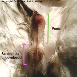
Image: The photograph above contains an image of a very infantile, underdeveloped tomcat scrotum. This cat was 4 years old and had all of the physical features (big head, thick skin, penis spines, heavy muscling, stinky urine) of a mature, entire male cat except for the fact that its scrotum was tiny! This animal was bilaterally cryptorchid with both of its testicles located within the abdominal region ahead of the bladder.
6. The presence of penis spines in the male cat:
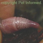 Entire male cats have a series of keratinised spines or spikes located on the head of their penis. These spines are designed to increase the friction of the tomcat's penis on the female cat's vagina during mating and, by doing so, induce the female cat to ovulate (female cats are induced ovulators which means that they do not ovulate their eggs from their ovaries unless they are stimulated to do so by the friction of mating). These spines are only maintained in the presence of testosterone, which means that they will be present on the penises of entire tomcats and cryptorchid tomcats, but they will not be present on the penises of neutered tomcats.
7. Examining the animal for scars:
Occasionally, you will come across cases whereby a previous vet has removed the scrotal testicles (i.e. castrated the animal) and/or unilaterally or bilaterally cryptorchid testicles (i.e. castrated the cryptorchid animal), but the current owner is unaware of this occurring. In these situations, it can be difficult to determine whether the animal was desexed or if it is, in fact, a bilaterally cryptorchid animal. You might get a clue that the animal has been desexed by looking for obvious past surgery scars in the regions of the prescrotum (just in front of the scrotum), inguinal region(s) and/or midline abdomen. The image below provides a visual reference to where these scar lines might be found.
Picture: The above image is a photograph taken of a cryptorchid dog immediately following surgery to locate a unilaterally-missing testicle. The penis is the small, dark, hairy mound in the centre of the image and the scrotal sac is the black lump on the left hand edge of the image. The dog's head is towards the right of this picture. The suture line indicated in pink is the site through which the scrotal testicle was removed during the castration. The suture line indicated in yellow is the incision made into the inguinal region where one might find a testicle lodged in the inguinal canal. The suture line indicated in white and running alongside the front edge of the penis is the incision made into the abdominal cavity where one might find an abdominally located testicle. Scars in any of these areas might indicate that a prior castration or cryptorchid-castration surgery has been performed.
8. Examining the ear for a desexing tattoo:
Most veterinary clinics place a distinctive desexing tattoo on the inside of the animal's ear flap (usually the left ear flap in Australia) to let other vets know that the animal (male or female) has been neutered. The tattoo is normally colored green or black and takes the form of a series of dots which are orientated in the shape of a circle with a line through it (it looks a bit like the no-smoking symbol). The presence of such a tattoo informs the veterinarian that the animal is desexed and not bilaterally cryptorchid.
9. Abdominal ultrasound:
It is sometimes possible for your veterinarian to locate bilaterally undescended testicles using ultrasound technology. Many clinics have access to diagnostic ultrasound these days and a skilled operator may be able to locate the undescended testicles. Note, however, that undescended testicles are often small and can be very difficult to find and that a negative result on ultrasound does not mean that the animal is not cryptorchid.
10. Testosterone assays:
Dogs that have been castrated normally have very low levels of testosterone in their blood because they lack the testicle organs which produce this hormone. Entire male dogs and cryptorchid dogs, however, tend to have much greater baseline levels of testosterone than neutered males do due to the presence of hormone secreting testicles. Measuring the level of testosterone in the blood of a suspect-cryptorchid dog can help to diagnose this condition. Cryptorchid males should have significantly higher levels of testosterone in their blood than desexed males.
Author's note: the blood testosterone levels of a bilaterally cryptorchid dog are usually less than the testosterone levels of a normally-descended entire male dog because the retained testicles are generally smaller in size than normally descended testicles. A small testicle makes less hormone. Despite this smaller size, the retained testes do produce significantly more testosterone than is found in a castrated male.
In cats, the situation is different. Studies have found that the blood testosterone levels of entire tomcats is often not all that significantly different to the blood testosterone levels of desexed toms. Consequently, a single measurement of blood testosterone in the cat is not always diagnostic for the presence or absence of testicles like it is in the dog. To use testosterone levels in the cat as a diagnostic aid for cryptorchidism, several steps have to occur:
1) The cat's baseline testosterone levels are measured - a blood test is performed (blood is taken) to measure the cat's normal, baseline testosterone production.
2) The cat is given an injection of either hCG (human chorionic gonadotrophin) or GnRH (gonadotrophin releasing hormone), both of which are designed to stimulate the testicles (if they are present) to release more testosterone.
3) One hour after the injection is given, the cat's post-stimulation testosterone levels are measured - a blood test is performed to measure the cat's testosterone levels following the injection of GnRH or hCG.
If the testosterone levels after stimulation with GnRH or hCG are greatly increased compared to the baseline testosterone values taken prior to hormonal stimulation, it is likely that the cat in question does have testicular tissue capable of responding to the stimulus and increasing testosterone output. Such a cat is likely to be a bilaterally or unilaterally cryptorchid animal. Neutered animals should show no significant increase in their blood testosterone levels after injection with hCG or GnRH because they have no testes to respond to the stimulus.
Author's note: I have not provided you with any specific blood testosterone measurements (figures) because these are not uniform across all of the different laboratories in the world. Your own local laboratories should have their own values of what they consider to be appropriate testosterone levels for an entire versus a neutered dog and so on.
11. Exploratory surgery:
Sometimes a diagnosis of bilateral cryptorchidism (as opposed to monorchism, anorchism or prior castration) can only be made by surgical exploration. In these situations, the animal is placed under a general anaesthetic and its entire abdomen, groin and scrotal region is shaved and surgically prepared. Because the missing testicles might be very small (retained testicles are often a lot smaller than scrotal testicles are) and could be located anywhere from the abdominal cavity, behind the kidney, through the inguinal canals to the prescrotal regions, the vet may have to make several incisions to find them. The vet might make an incision into the abdominal cavity (white line on the photograph below), only to discover that one or both of the retained testicles' spermatic cords (vas deferens and testicular blood vessels) disappears into the respective inguinal canal. The vet will then need to make a new incision into the appropriate right and/or left inguinal (groin) region/s (yellow line on the photograph below) to locate the missing testicle/s.
Picture: The above image is a photograph taken of a dog immediately following surgery to locate a unilaterally-missing testicle. The penis is the small, dark, hairy mound in the centre of the image and the scrotal sac is the black lump on the bottom edge of the image. The dog's head is towards the top of this picture. The suture line indicated in yellow is the incision made into the inguinal region where one might find an undescended testicle lodged in the inguinal canal. The suture line indicated in white and running alongside the front region of the penis is the incision made into the abdominal cavity where one might find an abdominally located undescended testicle.
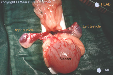
Photo: The picture above is a photograph of the inside of a cat's abdomen taken during a surgery to remove bilaterally undescended testicles. These testicles were located abdominally and had not entered the inguinal canals of the cat. To find the testicles, the veterinary surgeon had to incise into the cat's abdomen, find the cat's bladder (the large round structure labeled bladder) and then flip the bladder backwards towards the cat's tail. The testicular cords (vas deferens and testicular blood supply) are normally located just dorsal to the bladder neck, as seen in this image (the cat is lying on its back), and these cords can be followed until
the testicle/s are located. The testes could be located within the abdominal cavity (as seen in this animal) or they could have passed out through the pet's inguinal canals - either way, the testicle cords will trace out their path and find their location.
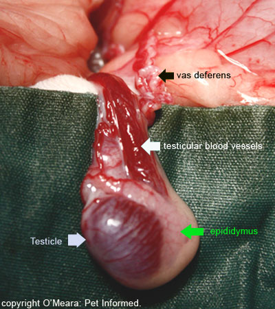
Image: This is a close-up view of one of the cat testicles. You can clearly see the testicle with its epididymus: this is the part that will be removed at surgery. You can also see the testicular blood vessels and the thick white worm-like structure that is the vas deferens. The bladder is the pink structure taking up the upper right hand side of the picture.
5. What are the medical implications and complications of cryptorchidism?
Cryptorchid testicles are prone to a number of significant and potentially life-threatening medical conditions that are not as commonly encountered in the correctly-descended testicle. These include: testicular torsion, testicular cancer and various testicular-cancer-related conditions such as male feminizing syndrome, oestrogen toxicity (this causes severe pancytopenia: a complete deficiency in the numbers of red blood cells, white blood cells and platelets in the animal's blood) and the hypersecretion of androgens such as testosterone. Each cryptorchid complication will be discussed in this section.
5a. Testicular torsion (twisted testicle condition):
Testicular torsion is the condition whereby the testis rotates on the end of its testicular cord (vas deferens and testicular blood supply), such that the vas deferens (also called the ductus deferens or spermatic cord) and testicular blood vessels become spiralled tightly around each other. This effectively results in the vas deferens strangling and cutting off the testicle's blood supply. The testis itself becomes starved of blood and is subsequently unable to receive life-giving nutrients and oxygen nor eliminate metabolic waste products such as lactic acid and carbon dioxide. The ultimate consequence of this is that the strangled testis initially becomes very swollen and enlarged and painful and then it gradually begins to die and rot within the animal.
Testicle torsion can occur within the scrotal sac (i.e. it can occur in properly descended testicles), however this is not very common because the testicle is typically held in a very tightly-fixed position within the scrotal pouch. The scrotal testis does not get much opportunity to move around, much less rotate about on its blood vessel and vas deferens attachments. When the condition does occur in a scrotal testicle, the pet develops a very hot, swollen, painful, enlarged testicle and scrotum. The animal will be reluctant to sit; it will seem painful; it will not allow the testicle to be touched and it will tend to constantly lick and bother at the painful, swollen region. Left long enough, the scrotum may become necrotic (rot) and turn black and the animal will become sick. Treatment involves removal of the affected testicle (i.e. castration of that damaged testicle) and the prognosis is normally good.
Whilst testicular torsion is very uncommon in the scrotal testicle, it is significantly more common in an undescended testis, particularly an undescended, abdominally-located testis. Abdominal testicles are not tightly fixed into position (see the image of the cryptorchid cat testes below) and are, instead, free to float about within the abdominal cavity on their long vascular and spermatic duct cords. They therefore have much more chance than scrotal testes do of spinning about and twisting on their testicular attachments, leading to testicular torsion and strangulation. The chances of a cryptorchid animal developing a testicle torsion is particularly high if the retained testicle has developed a cancer (tumour) of any kind. Enlarged, undescended testicles with large tumors growing off them are very likely to become unbalanced and rotate about on their blood vessel attachments, particularly when the animal runs around (big tumour-testicles will tend to bounce about in the belly much more than small testicles will).
When torsion does occur in an abdominal testicle, the animal will initially present with signs of acute abdominal pain and a reluctance to move. The animal might be inappetant (not willing to eat) and lethargic and exhibit other signs of internal discomfort (e.g. panting, teeth grinding, pacing, restlessness, vomiting, crying when touched). If the condition goes undiagnosed, the torsed abdominal testicle will rot, resulting in peritonitis (severe inflammation of the abdominal cavity); ascites (an abdomen full of fluid); vomiting; severe shock (collapse, high heart rate, pale gums, cold extremities and so on) and even death. Treatment involves supportive care (fluids, antibiotics, treatment for shock) and immediate surgical removal of the affected testicle. The prognosis is good if the condition is caught early, but guarded to poor if the condition is diagnosed late and severe peritonitis and shock has set in.
Image: This is the picture of the cat with the bilaterally cryptorchid abdominally-located testicles. Notice how long the blood vessels and spermatic ducts are, which supply these testicles. There is plenty of scope for these testes to rotate about on their testicular cord attachments, resulting in torsion of the testis.
5b. Testicular cancer (testicle cancer):
Canine testicles, even normally-positioned scrotal testicles, have a high chance of developing testicular cancer, with a risk that increases the longer the dog in question remains uncastrated. The prevention of canine testicular cancer is one of the many reasons why veterinarians promote the neutering of male, non-breeding, pet dogs. In the case of undescended testicles, particularly intra-abdominal testes, the risk of the undescended testicle or testicles developing cancerous changes is higher still and is the main reason why veterinarians advocate the surgical removal of undescended testes in the dog.
Undescended testicles are prone to many types of cancer, the most common and well-known being: Sertoli Cell Tumours, Seminomas and Interstitial Cell (Leydig cell) Tumours. Other forms of cancer are also possible, but less common. Sertoli Cell Tumours are cancers that result from the unregulated replication of mutated testicular Sertoli Cells (Sertoli Cells are cells that line the spermatic ductules (small ducts) within the testis and which assist the sperm to mature into fertile, swimming forms prior to leaving the testicle). Seminomas are cancers that result from the unregulated replication of mutated testicular sperm-production cells. Interstitial Cell Tumours (also called Leydig Cell Tumours) are cancers that result from the uncontrolled replication of mutated testicular Interstitial Cells (Leydig Cells) whose normal role in the body is the production of testosterone. Sertoli Cell Tumours and Seminomas can become quite large in size (around baseball size) and both have the potential to spread to other organs (metastasize), with Sertoli Cell Tumours more likely to spread than Seminomas. Interstitial Cell Tumours normally remain small and benign and very rarely spread to other organs.
Testicular tumors can produce a wide range of clinical effects on the affected animal. The testicle tumor itself can produce symptoms of abdominal pain as it grows and stretches the nerve-rich testicular capsule. Large tumors can burst, causing severe abdominal bleeding; they can compress and invade body structures located nearby (e.g. other organs), resulting in dysfunction of these organs and resultant illness; they can even outgrow their own blood supplies, resulting in sections of the testis tumor dying off and rotting and even becoming infected (bacteria like to lodge in dead tissues and make abscesses) and the animal becoming very febrile and sick (septic shock) as a result of blood poisoning with bacteria and toxins. Some types of testis tumors spread throughout the body, invading and destroying many tissues: a terminal condition that eventually results in severe weight loss, organ dysfunction and death. Some testicular tumours may even secrete excessive quantities of sex hormones such as oestrogen and testosterone, a situation that results in the affected animal displaying a wide range of severe hormone-induced disease symptoms. For example: Sertoli Tumours are well known for secreting excessive amounts of oestrogen, a female hormone that, in excess, can produce severe pancytopenia and male feminizing syndrome (see sections 5c and 5d) in animals. Some Leydig Cell Tumours (Interstitial Cell Tumours) secrete excessive amounts of testosterone, resulting in greatly masculinised features; possible damage to and dysfunction of organs such as the liver and prostate and an increased likelihood of medical conditions such as perianal adenoma and prostatic hyperplasia occurring (see section 5e). Some animals with cancer of the testis will show minimal signs of disease and may only present for infertility. This infertility may be a result of compression of sperm-producing tissues by the testicle cancer; imbalanced testosterone or estrogen production or inflammatory breakdown of the blood-testis barrier and exposure of the 'foreign' sperm to the male animal's immune system (which then subsequently attacks all sperm rendering the animal infertile).
The treatment for testicular cancer really depends on whether or not the cancer has spread to other organs and whether or not other complications of the cancer are apparent (e.g. septic shock, estrogen toxicity). Treatment (and prevention) of testicular cancer involves removal of the testicles (castration). Because it is well-known for both testicles to develop cancerous changes at the same time or one after the other, it is normally recommended that both testicles be removed, not just the cancerous one.
Author's note: Testicular cancer is rare in the cat compared to the dog. It is not common for a normally-descended or cryptorchid cat to develop testicular cancer, however, it can occasionally occur. Castration of cryptorchid cats is still recommended.
5c. Male feminising syndrome:
Certain testicular cancers (particularly Sertoli Cell Tumours) are well-known for secreting large amounts of a "female hormone" called estrogen (also spelled oestrogen). Estrogen is the hormone that makes ladies look and act like ladies: it causes bitches and queens to have large mammaries (teats) compared to male animals; it makes female animals attractive to males; it makes female animals receptive to males; it affects the growth of the female hair coat and so on. Excessive oestrogen production in a male dog or cat with a Sertoli Cell Tumour (or other estrogen secreting mass) can have a similar effect to that seen in the female animal. The male animal will become "feminized" by the oestrogen. It will develop larger mammary glands; it will become attractive to other males of its species; feminised male dogs may cease cocking their legs to urinate and instead squat like females and some males will even have a reduced sex-drive (libido) as a result of estrogen suppression of testicular testosterone production. The normal, non-cancerous testicle will often atrophy (shrink in size) as a result of this reduced testosterone production. Many estrogenised dogs will show significant changes to their skin and hair coat (this is often the first thing an owner will notice - the first outward sign of estrogen-producing cancer). The animal's skin may darken with increased skin pigmentation (it will often turn black) and the animal will often become symmetrically bald, initially losing the hair from around the anus and genital regions, before gradually losing all of the coat on the belly, back, chest and neck (usually, only the fur on the head and legs remains). The skin condition is not itchy. If the oestrogenising condition is allowed to progress and remain untreated, the estrogen can cause significant changes to the structure of the male's prostate (estrogen causes squamous metaplasia - a flattening and sloughing of the cells lining the prostate gland), resulting in prostate dysfunction, infertility, prostate pain, urination pain, prostatic inflammation (prostatitis) and even secondary prostate infections and abscesses. Prolonged, excessive oestrogen production can also result in a life-threatening complication called estrogen toxicity, which manifests as nasty bone marrow hypoplasia and pancytopenia (section 5d).
Treatment of male feminising syndrome involves removal of the estrogen secreting tumour or tumours: i.e. it involves castration of both testicles (testicle removal). Extra therapy and supportive care (see section 5d) may also be required if the bone marrow and blood cells have been affected by oestrogen toxicity. With successful removal of the oestrogen secreting tumour, the symptoms of male feminising syndrome should resolve, however, it may well take many months (4-6 months) for the haircoat to regrow if it has been lost and for the prostate's structure to normalise. Failure of the feminising symptoms to resolve following castration suggests that the tumour has spread to other organs and that there are still foci of estrogen secreting cells lurking about the body.
Author's note: Undescended, cryptorchid testicles that remain within the abdominal cavity are particularly prone to developing Sertoli Cell Tumours and male feminising syndrome (more so than inguinal canal or prescrotal cryptorchid testes or normal scrotal testes).
5d. Bone marrow hypoplasia and pancytopenia - estrogen toxicity:
Excessive estrogen production, as can occur in male animals with certain testicular tumours or female animals with estrogen-secreting ovarian tumours or estrogen-secretory ovarian follicular cysts, can cause the feminising syndrome described in section 5c. In its severe form, it can also produce signs of estrogen toxicity, resulting in severe, often life-threatening effects on the animal's bone marrow.
The bone marrow of the animal is the place where most of the animal's blood cells are produced: the red blood cells, white blood cells and platelets. The red blood cells are responsible for carrying oxygen to the animal's body tissues - without them, the tissues of the body become starved of oxygen and start to die. The white blood cells are responsible for fighting infectious diseases and tumours and other foreign invaders - without them, the body becomes prone to infection by bacteria, fungi, viruses, other micro-organisms and even cancer. The platelets are responsible for helping the animal's blood to clot - without them, the animal is likely to suffer from severe, uncontrollable hemorrhage every time it receives a tiny little bump or scratch.
Excessive amounts of estrogen are highly toxic to the animal's bone marrow. The estrogen causes the bone marrow precursor cells (juvenile blood cells located inside of the bone marrow, which are responsible for creating all of the mature blood cell lines) to die off. This bone marrow cell death, termed bone marrow hypoplasia, results in a lack of production of new red blood cells, white blood cells and platelets. The consequence of this is that, as the old cells in the blood circulation (red cells, white cells and platelets) get consumed and/or die off with age, there are no new cells being made by the bone marrow to replace their numbers. As a result, all three blood cell lines start to decrease in population.
The animal eventually becomes very sick as a result of loss of all three of these blood cell lines. As a result of red blood cell deficiency (anaemia), the animal becomes pale and weak and collapsed (shocky), with a high heart rate and heavy respiratory pattern. Without urgent treatment (e.g. blood transfusion), a severely anemic animal may die as a direct consequence of insufficient body-tissue oxygenation. A complete deficiency of white blood cells causes the animal to be prone to developing infections (bacterial, fungal, viral and so on) in multiple body regions including the intestines, lungs, skin and bladder. This produces a vast range of symptoms, depending on the body region/s infected, including: vomiting, diarrhea, colitis, fever, lethargy, pneumonia, coughing, pussy nasal discharges, trouble breathing, skin infections, itchy skin, abscesses, urinary tract infections, peritonitis and even life-threatening complications such as bacteraemia (bacteria in the bloodstream), blood poisoning (termed toxaemia - bacterial poisons in the blood), septic shock and death. As a result of blood platelet deficiency, the animal is prone to excessive bruising and prolonged bleeding and may spontaneously hemorrhage into any region of its body. Severe shock and even death may result from rapid, acute, severe blood loss.
Estrogen toxicity is diagnosed on the basis of:
1) a complete blood count (CBC): examination of the animal's blood under a microscope reveals a complete lack of most of the blood cell lines;
2) a bone marrow aspirate: examination of the bone marrow samples under a microscope reveals a lack of bone marrow precursor cells - i.e. bone marrow hypoplasia - and
3) a blood estrogen assay: the animal's blood contains excessive levels of estrogen.
Treatment of the oestrogen toxicity condition involves removal of the source of estrogen production (i.e. removal of the tumourous testicles) as well as expensive, intensive supportive care. The severely oestrogen affected animal may spend weeks to months in hospital; it may require repeated blood transfusions and platelet-rich plasma transfusions; it will need loads of broad-spectrum antibiotics (to fight off infection while the white blood cells are recovering) and it may even require expensive, difficult-to-obtain bone marrow stimulating drugs such as erythropoietin (epo) and granulocyte-colony-stimulating-factor (G-CSF). A bone marrow transplant may also need to be considered.
The prognosis for recovery is very poor with severe bone marrow hypoplasia and pancytopenia. Recovery, if it does occur, will often take up to 6 weeks to become evident (the animal may need to be hospitalized the whole time). Some animals will never recover, regardless of the treatment given. Therapy is not expected to be successful if the estrogen-secreting tumour has spread to other organs and there are still foci of estrogen secreting cancer cells lurking about in the body.
5e. Excessive testosterone production:
Leydig Cells, otherwise known as Interstitial Cells, are the testicular cells responsible for secreting the male hormone: testosterone. Occasionally, Leydig Cell Tumours can arise in normally descended or cryptorchid testicles which are "functional". This means that they secrete excessive quantities of testosterone into the animal's bloodstream. Because testosterone is the hormone responsible for making males look and act like males, excessive amounts of testosterone produced by testicular tumours can cause the affected animal to look and behave in an excessively masculine manner. The animal will become heavily muscular and develop a masculine appearance. The animal will act like an entire male, showing increased interest in females of the same species; increased sexual drive; increased inter-male aggression and, potentially, increased dominance and aggression in general. Certain testosterone-enhanced medical conditions and diseases may also appear in testosterone increased animals, including prostatic enlargement (prostate hyperplasia), prostatitis, prostate dysfunction, prostatic abscessation and perianal tumours (small cancers that occur around the anuses of male dogs that are thought to be induced by testosterone secretion in entire male animals). As before, treatment of the condition requires removal of the testosterone secreting cells and, therefore, castration of both testes (testicle removal).
6. What are the behavioural implications of cryptorchidism?
As shall be discussed in sections 7 and 10, the cryptorchid testicle or testicles are normally infertile: a result of core-body-heat-induced destruction of the spermatogenic cells that are responsible for producing the sperm within the testis. Despite being infertile, these retained testicles are still capable of producing moderate to normal amounts of blood testosterone: the hormone that makes male animals look and act like male animals. Unilaterally or bilaterally cryptorchid animals are therefore very likely to exhibit the kinds of "male" testosterone-dependent behaviours normally attributed to an entire animal. Cryptorchid tomcats and dogs may be more aggressive and more dominant and more prone to male-to-male aggression (inter-male aggression) than neutered animals: i.e. they act like bossy entire males. They will tend to exhibit sexualised behaviours including: aroused interest in females of their own species; mounting of females (particularly in-heat, estrous females) and complete erection of the penis when excited. They may be more prone to displaying masculine territorial behaviours such as the guarding of resources (food, bones, territory, companion people and pets and so on) and the marking of territory with urine and faeces (e.g. cryptorchid tomcats may exhibit urine spraying in the house; male dogs will cock their leg to urinate on vertical surfaces). Additionally, cryptorchid male pets, just like other entire male animals, will be more likely than neutered animals to leave their yards and roam the countryside looking for females and trouble. Roaming is a troublesome habit because it puts other animals (wildlife and other pets) and humans at risk of harm from your animal and it puts the roaming pet at risk from all manner of dangers including: predation by other animals, cruelty by humans, poisoning, envenomation (e.g. snake bite) and motor vehicle strikes.
7. What are the economic and showing implications of cryptorchidism?
Although cryptorchidism sounds like an innocent enough condition to diagnose and treat (e.g. via castration), it does have a number of significant implications with regard to the commercialised breeding, showing and selling of animals.
Loss of a potentially valuable breeding animal:
Cryptorchidism is considered to be an inherited condition that can be passed on from affected parents to their offspring. The abnormal gene (genes?) that produce cryptorchidism can be carried and passed on by both dam and sire (mother and father animal), however, only the male offspring which inherit the right combinations of cryptorchid genes will actually display the condition (only males have testicles and thus only males can be cryptorchid). The inheritance of cryptorchidism is discussed in detail in section 10. Since the cryptorchid condition can be passed on from affected parents to their offspring, it is considered unethical to deliberately breed from known cryptorchid males because this will most likely result in the passing on of a significant breed flaw (undescended testes) to the next generation and result in affected offspring potentially developing life-threatening medical complications such as pancytopenia and testicular cancer. The consequence of this is that, no matter how good the affected male animal is otherwise (i.e. it might otherwise have excellent show lineage/pedigree, temperament, breed conformation and features), cryptorchidism essentially rules out that valuable animal as a breeding animal.
Infertility:
Bilaterally undescended testes are normally infertile because of heat-induced damage to the sperm-producing cells of the testicle. The bilateral cryptorchid male animal is therefore infertile.
Unilaterally cryptorchid animals are normally still fertile because of normal sperm production by the fully-descended testicle.
Although it is possible (though unethical) for cryptorchid testicles to be surgically transferred from the abdominal cavity or inguinal region to the scrotal region, it is likely that many of these relocated testicles will have already suffered irreparable heat damage and, therefore, remain infertile and incapable of producing sperm despite their new scrotal location.
Loss of lineages:
If a cryptorchidism affected dog or cat or horse is the 'last of its line' (i.e. the last pup in a long line of pedigree breeding dogs), a breeder's choice not to mate that animal and, therefore, not pass on its valuable breed genetics (so as to not pass on the undesirable cryptorchid genes) will essentially spell the end of that breeding lineage.
Loss of the affected animal's parents and siblings from the breeding pool:
When a cryptorchid animal (or animals) appears in a litter, the implication of this is that both of the parents, male and female, must have been carriers of the defective cryptorchid genes, even if the sire did not show any outward sign of cryptorchidism himself. This is due to the recessive way in which the cryptorchid gene(s) is/are thought to be passed down the generations (see section 10c for full details). Additionally, because the parent animals are both carriers of the defect, there is every likelihood that some of the other offspring in the litter (the siblings of the affected animals) may also be carriers of the abnormal genes. Therefore, if control and eradication of the cryptorchidism condition is to occur in a breeding population, the parents of the affected animals, as well as any siblings of the affected animals, should not be bred from. This constitutes a significant loss of breeding potential from that one kennel or cattery.
Poor show-ring performance:
Cryptorchidism is considered to be a major breed fault in most show-ring animals and animals detected with the condition are normally left unplaced and out of the ribbons. Failure to place in dog or cat shows for any reason can have a significant effect on the affected owner's business if their business is the production of high quality, show quality animals. Breeders whose pets do not place well in shows will be less likely to be able to sell their animals to other breeders or show participants for a high price (after all, show performance and pedigree are everything). Should the reason for non-placement of the male animal become well known (breeders talk!), the sales of any animals from the cryptorchidism-affected kennel to other breeders and show types may well cease altogether. No reputable animal breeder or show-animal producer wants to run the risk of introducing bad genes (e.g. cryptorchid genes) into his or her kennel.
Loss of reputation:
Reputation is everything in the dog and cat showing and breeding world. Any kennel that becomes well known for selling animals which are cryptorchid or likely to throw cryptorchid pups and kittens may become black-listed by the local breeding and showing community. This can result in consistently poor results at local and regional shows and cause sales of breeding-quality male and female animals to dry up.
Medical costs:
As mentioned previously (section 5), there are significant medical problems that can arise as a direct result of cryptorchidism. Some of these conditions can become quite life-threatening and very costly to treat with regard to the veterinary care that might be needed.
The medical costs of cryptorchidism are not limited to the costs of treating the cryptorchidism related complications alone. They also include the costs that can be incurred when animals come to grief during testosterone-fuelled roaming and wandering activities. Roaming animals can be poisoned, hit by cars, bitten by snakes and attacked by other animals: all of which can result in complications (e.g. broken bones, abscesses, organ failure) that may require expensive veterinary treatment.
At the very least, you can expect the costs of desexing a young, healthy, cryptorchid dog or cat to be higher than the costs of desexing a male with normal scrotal anatomy. It takes much longer to find and remove the testicles of a cryptorchid dog or cat than it does to remove the testes of a normal male. This increase in costs will be particularly noticeable to those of you attempting to desex cryptorchid horses and livestock animals - finding and removing the undescended testicles of a large animal is incredibly difficult and time-consuming and expensive to do.
For those of you hoping to fall back on your pet insurance, think again. It is unlikely that pet insurance companies will insure pets against medical complications that arise as a result of cryptorchidism. Most insurance companies will not protect pets against congenital problems (birth defects) or pre-existing problems and nor will they protect animals against proven inherited conditions. Cryptorchidism is all of these and, as a result, pet insurance companies will be unlikely to cover pets against cryptorchidism related illnesses and complications.
8. Should I buy (purchase) an animal if it is cryptorchid?
Having read about cryptorchidism and its behavioural, medical and economic consequences thus far, you might have come to the conclusion that cryptorchidism is a terrible affliction and that one should never purchase a dog or cat affected with the condition. This is not completely true. Whether or not you purchase a male animal affected with cryptorchidism really depends on the age of the animal and what it is that you want the animal for. Do you want that animal to be a breeding or showing animal or do you merely want that animal to be a loving family pet?
If all you are after in your choice of dog or cat is a nice family pet, then a cryptorchid male is a fine choice, provided you get that dog or cat desexed early and prior to the onset of any unpleasant medical conditions (tumours etc). Once that cryptorchid animal has been desexed, it should essentially be the same as any other neutered male with regards to its medical and behavioural health. Provided the animal receives the right training and treatment, this neutered cryptorchid male should have the same chance as any other neutered animal of becoming a good and loving family companion pet.
Author's note: I would exercise some caution if considering taking on a cryptorchid family pet (especially a dog) that is over 6 years of age. The chances of a cryptorchid dog over the age of six years having some form of testicular cancer is moderately high. You should request that the prospective pet animal be desexed by a veterinarian and its testes assessed for testicular cancer prior to taking on the animal. If the animal is desexed and given the all-clear by the vet, then there is no reason why the animal should not make a good family pet.
If you are seeking a dog or cat for breeding or showing purposes, then a cryptorchid animal is not for you, for all of the economic and reputational reasons indicated in section 7. You do not want to risk bringing known cryptorchid genes into a cryptorchid-free breeding population. I would even go so far as to say that you should not purchase any dogs or cats (cryptorchid or otherwise) from a breeder who would offer you a cryptorchid breeding animal for sale. Not only is such a breeder likely to have many cryptorchidism carrier females and males in his or her kennels, but such a breeder obviously does not have any breeding ethics (who knows what other genetic defects might be running rampant in that person's facilities besides the cryptorchidism).
9. Desexing (neutering) a cryptorchid male (i.e. surgical testicle removal):
9a) Why should you get a cryptorchid animal neutered (castrated)?
Having read up to this point, particularly sections 5, 6 and 7, it should now be self evident why the castration of cryptorchid dogs and cats is highly recommended by most veterinarians. Cryptorchid cats and dogs are prone to a number of potentially life-threatening and/or problematic medical conditions that can be prevented through early castration and removal of the undescended testicle/s. Early castration of cryptorchid and, for that matter, entire male dogs and cats can also reduce and prevent the onset of various testosterone-related behavioural problems including: urine spraying in tomcats, aggression, dominance, inter-male aggression, fighting, roaming and escaping from yards and hypersexual behaviours. The final reason why the neutering of cryptorchid animals is highly recommended is because the condition is heritable and can be passed on from the affected animal to its offspring. Castration prevents the animal from breeding and passing on defective genes.
9b) How do vets desex (castrate) a cryptorchid animal?
 The castration of a cryptorchid animal normally occurs via a similar surgical process to that already described in the sections (section 4a and 4b) discussing the surgical exploration and subsequent diagnosis of unilaterally or bilaterally cryptorchid testes.
The animal is placed under a general anaesthetic and its entire abdomen, groin and scrotal region is shaved and surgically prepared. Because the missing testicle or testicles might be very small (retained testicles are often a lot smaller than scrotal testicles are) and could be located anywhere from the abdominal cavity, behind the kidney, through the inguinal canal to the prescrotal region, the vet may have to make several incisions to find the cryptorchid testis or testes. The vet might initially make an incision into the pet's abdominal cavity (white line on the photograph below), only to discover that the retained testicle's or testicles' spermatic cord/s (vas deferens and testicular blood vessels) disappear out into the respective inguinal canal/s. The vet will then need to make a new incision into the appropriate right and/or left inguinal (groin) region(s) (yellow line on the photograph below) to locate the missing testicle/s.
Picture: The above image is a photograph taken of a dog immediately following surgery to locate and remove a unilaterally-missing testicle. The penis is the small, dark, hairy mound in the centre of the image and the scrotal sac is the black lump on the bottom edge of the image. The dog's head is towards the top of this picture. The suture line indicated in yellow is the incision made into the inguinal region where one might find and remove a testicle lodged in the inguinal canal. The suture line indicated in white and running alongside the front part of the dog's penis is the incision made into the abdominal cavity where one might discover and remove an abdominally located testicle.
Photo: The picture above is a photographic image of the inside of a cryptorchid cat's abdomen, taken during surgery to remove bilaterally undescended testicles. These testes were located abdominally and had not yet entered the inguinal canals of the cat. To find the testicles, the veterinary surgeon had to incise into the cat's abdomen, find the cat's bladder (the large round structure labeled bladder) and then flip the bladder backwards towards the cat's tail. The testicular cords (vas deferens and testicular blood vessels) are normally located just dorsal to the bladder neck (the cat is lying on its back). All the surgeon needs to do is follow these cords until the testicle/s and epididymi are located. These retained testicles could be located within the animal's abdominal cavity, as seen here, or they could have passed out through the cat's inguinal canals - either way, the testicle cords will trace their path and find their location.
Sometimes the retained testicle is very obvious, sitting outside of the abdominal cavity, forming a pronounced lump or bulge just under the skin in the groin region (see image below). In these cases, the veterinary surgeon will not need to incise into the animal's abdomen to find and neuter the cryptorchid testicle. The vet will simply make an incision through the groin skin, directly over the retained testicle, and remove the testicle through the hole in the skin.
Images: These photographs have appeared previously on this site and clearly show an undescended testicle located in this Pomeranian dog's right inguinal region. In this case, the veterinarian will not need to enter the animal's abdominal cavity surgically. All the vet will have to do is incise over the testicle lump (indicated with the green arrows) and remove the testis via the skin.
What parts get removed during the castration of a cryptorchid animal?
The parts of the testicle that get removed during the castration of a cryptorchid animal are the same as those that get removed during the normal castration of a fully-descended male dog or cat: the testis and epididymus. Basically, the parts removed are those responsible for producing sperm and testosterone: the causes of all male-related problems for pet owners. During castration of a normal or retained testicle, the testicle is exteriorised (raised from the body or scrotal sac); the testicular blood vessels and spermatic cord (vas deferens) are tied off with surgical thread (so that they do not hemorrhage when cut) and the testicular blood vessels and cord are chopped off above the level of the surgical thread, thereby removing the testis and epididymus. The image below is of the same bilaterally cryptorchid cat pictured before. I have marked on the image in green lines where the testicular cords will be ligated (tied off) and incised so that the testes and epididymides can be removed and discarded.
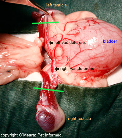
Image: Bilaterally cryptorchid cat undergoing desexing. The green lines indicate where the testicular cords (vas deferens and testicular blood supply) will be tied off and incised.
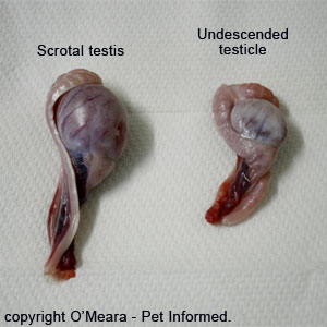
Image: Image of dog testicles that were removed at surgery. This picture clearly shows the parts of the testicles that are removed during castration. Notice how small the cryptorchid (undescended) dog testis is compared to the scrotal testis. Atrophy of the testes is a common feature of cryptorchid testicles.
9c) Can the vet just remove the internal testicle and leave the descended testicle intact?
In the case of a dog or cat with unilateral cryptorchidism, just removing the cryptorchid testis and leaving the descended testis intact will go some way towards preventing most of the medical complications of cryptorchidism, particularly if the surgery to remove the retained testicle is performed early on in life (i.e. before the onset of cancer). It is not advisable, however, to just remove the single retained testicle and leave the descended one intact. The main reason for this is that the cryptorchidism condition is hereditary. Leaving the scrotal testicle (which is fertile) intact will enable the cryptorchid animal to father young which will carry the defective cryptorchidism gene/s. In addition to this, if the animal in question is old, there is every possibility that the cryptorchid testis and possibly even the descended testis may contain cancerous changes. Leaving the descended testis intact may pose a risk to that animal should the descended testicle contain cancer.
9d) Are there drugs, medications or surgical procedures available to make the retained testicle descend (i.e. cryptorchidism repair) and what are the ethical and medical implications of this?
Despite the fact that cryptorchidism is an undesirable hereditary trait, best eliminated through selective castration and non-breeding, there are still many breeders and show-types who refuse to discard a cryptorchid show dog or stud dog and who want their veterinarian to prescribe treatments to descend their animal's testicle or testicles so that they can hide the cryptorchidism fault from potential stud clients, show-judges and dog buyers. Options that have been trialled, with variable success, include both medical options (hormones, drugs) and surgical options.
Medical options:
Most veterinary texts that discuss the matter of drug or hormone-assisted testicular descension suggest the attempt to be, as a general rule, unrewarding. Most veterinary texts say that there is no highly-effective medical treatment available for treating cryptorchidism.
The only report of successful medicine-based induction of testicular descension that I have located comes from the Feldman and Nelson book of Canine and Feline Endocrinology and Reproduction (reference number - 2). This reference indicates that some success in inducing canine (feline?) testicular descension might be achieved by administering either hCG (human chorionic gonadotrophin) or GnRH (gonadotrophin releasing hormone), both of which act on descended or undescended testicular Leydig Cells (Interstitial Cells) to induce them to produce increased quantities of testosterone. The reason these treatments are thought to have some merit is because normal testicular descent (gubernaculum regression) is thought to be mediated by blood testosterone levels. Because cryptorchid testicles usually secrete slightly less testosterone than normally-descended testicles, it is thought that some benefit might be gained by hormonally inducing increased amounts of testosterone to be released.
GnRH: Two doses of GnRH are usually administered at an interval of 1 week apart.
hCG: Four doses of hCG are usually administered over a 2 week period.
There does not seem to be any benefit gained by giving artificial testosterone itself, which is why the GnRH or hCG is used instead.
According to the reference (reference 2), these drugs have only been found to be somewhat successful in the induction of canine testicular descension if they are given to puppies under 16 weeks of age. Results are very variable and higher success rates seem to be found in dogs whose undescended testicle or testicles are located outside of the abdominal cavity already. Whether the resultant successful testicular descension that occurs is a result of the drugs themselves or a result of the fact that these testicles might have descended anyway is unknown.
Surgical options:
The surgical relocation of an abdominally, inguinally or prescrotally located retained testicle into the scrotal sac is considered to be both a highly-complex and unethical procedure. It is, however, a procedure that is offered to dog breeders and showers by some veterinarians. The surgical procedure is not without potential complications, including the death of the translocated testicle through damage or occlusion of the testicular blood supplies, and there is always the likelihood that the relocated testicle/s will remain infertile, having been previously located at body temperatures warm enough to cause permanent damage to the spermatogenic cells of the testis.
The artificial induction of testicular descent, whether this be through medical or surgical processes, must always be considered questionable from an ethical viewpoint. The condition is a hereditary defect with medical implications for the affected animal and any of its affected offspring. The deliberate breeding of any animal with such hereditary defects should be frowned upon as unethical because of the detriment to the breed's quality overall and because of the suffering and pain (cancer, testicular torsion etc.) that might be experienced by afflicted dogs further down the line.
The practise of inducing testicular descent artificially should also be considered unethical from a business viewpoint too. The whole reason why breeders and showers of dogs and cats seek to get their animal's testicles corrected is to deceive show judges, prospective breeder clients and prospective animal buyers into thinking that their animal is genitally and genetically sound. This results in more ribbons at the show, more stud-dog sire demand and more pups sold to the deceived buyer, but in doing so, it essentially robs the ethical showers of their deserved success and defrauds the owners of breeding bitches and queens who subsequently have their females mated to that prize-winning, but defective stud.
10. Breeding from a cryptorchid animal:
The following section contains information about the fertility and libido of cryptorchid males and answers questions about the genetics and heritability of the condition, as well as the ethical implications of breeding an animal with cryptorchidism.
10a) Are cryptorchid males fertile and able to breed?
Whether or not a cryptorchid animal is able to breed successfully depends on whether or not that animal has at least one normally-descended testicle. Only fully descended, scrotally-contained testicles are cool enough for viable sperm to be produced successfully.
Animals with bilaterally undescended testes are generally infertile and unable to father young.
Animals with unilaterally undescended testes are generally fertile and able to father young. This is because they have one normally-descended scrotal testicle.
10b) Do cryptorchid animals exhibit typical male behavior and libido?
As discussed in section 6, cryptorchid testicles are still capable of producing moderate to normal levels of blood testosterone: the hormone that makes male animals look and act like male animals. Unilaterally or bilaterally cryptorchid animals are therefore able to exhibit the kinds of "male" testosterone-dependent behaviours normally attributed to an entire animal. Cryptorchid animals do exhibit typical entire male behaviour: they are usually more aggressive, more dominant, more territorial and more prone to inter-male aggression than neutered animals. Cryptorchid animals do tend to have normal libido and they will exhibit masculine sexual behaviours including: aroused interest in females of their species; mounting of females (particularly in-heat, estrous females); mating of females and complete erection of the penis when excited.
10c) Is cryptorchidism hereditary? What is the mode of inheritance?
As mentioned in previous sections, cryptorchidism is a hereditary condition that can be passed on from affected sires and dams to their male and female young (only the male offspring can actually display the disorder because female offspring do not have testicles, however, affected female offspring can carry the genes through to the next generation thereby passing the disorder on).
The specific gene or genes that produce cryptorchidism in the dog and cat have not yet been positively identified as far as I know (i.e. there is not yet a genetic screening test that can be performed to detect cryptorchidism-affected and cryptorchidism-gene-carrier animals). There is even still conjecture about whether just one defective gene or many defective genes are responsible for the defect.
What has been determined to some extent is the mode of inheritance in the dog. I use the words "to some extent" because even the mode of inheritance of canine cryptorchidism has been difficult to establish for certain. At the current time, cryptorchidism is thought to have an
autosomal recessive mode of inheritance in the dog. The mode of inheritance has not been so well studied and determined in the cat, but is thought to be similar to the dog.
A primer on basic genetics:
Before I can discuss the implications and consequences of an
autosomal recessive mode of inheritance, I need to give you a basic overview of simple genetic principles.
All human and animal cells contain DNA. DNA is a long sequence of four different kinds of molecules (A,T,C,G) which can be arranged into millions of different combinations of varying lengths to code for all of the genes in a body. The information contained in each DNA gene sequence is used by the cell as a template or set of instructions by which that cell is able to manufacture certain important proteins and molecules vital to cellular function, replication and survival. Genes code for pretty much everything in the body: from the production of vital hormones (e.g. insulin, growth hormone, cortisol, testosterone) to the production of enzymes used in important cellular chemical reactions to the production of proteins and compounds that imbue humans and animals with their important body traits (e.g. eye colour, hair colour, coat length, coat softness, body size, baldness and so on).
Keeping to this webpage's main topic, there is a gene or a number of genes responsible for making the testicle descend into its normal scrotal location in early life. This might be a gene with a role in testosterone production - after all, sufficient testosterone production is thought to assist normal testicular descent (hence the medical use of GnRH or hCG to try to correct cryptorchidism). Alternatively, it might even be a gene with a role in normal gubernaculum development (e.g. a gene responsible for making normal gubernaculum contractile proteins) - after all, a normally-regressing gubernaculum is required in order for the testicle to descend. Correct testicular descent could and is likely to be a combination of many different genes all working together. A defect in any one of these genes, which prevents the cells from working correctly and making the right gene-coded products (e.g. prevents them from producing the right molecular forms of testosterone; prevents them from producing enough testosterone; prevents them from producing normal gubernacular contractile proteins ...) could result in a failure of normal testicular descent and, therefore, cryptorchidism.
To make matters more complex, every cell in the body does not merely contain just one set of all of the genes, but two. One set from the animal's mother and a second set from the animal's father. This means that, for every single gene-coded trait that exists in a body, there are two possible parental gene combinations that could code for it. This is where the issue of inheritance comes in - animals and humans inherit genes and, therefore, traits from
both of their parents. For example, an animal might inherit a gene coding for blue eyes from its father and a gene coding for brown eyes from its mother. Either gene could be switched on in that animal, giving the offspring animal blue eyes or brown eyes. Which color
actually gets expressed by the young animal's eyes really depends on the nature of the different genes themselves (the gene for blue versus the gene for brown) and which is the more
dominant gene. If the gene for blue eyes is a dominant gene, then the mother's gene for brown eyes will not be expressed in that young animal (the non-expressed brown eye gene is termed a
recessive gene) and it will display blue eyes. The young animal will be a
carrier of the brown eye gene (it might even pass the brown eye gene onto its own offspring, giving them brown eyes) but it will not have brown eyes itself because of the overpowering dominance of the blue gene.
In the case of cryptorchidism, the defective gene (or genes) that produce cryptorchidism are thought to be recessive to the
normal versions of the same gene (the genes that produce normal testicular descent). Therefore, if a young male inherits a normal version of the gene (normal testicular descent gene) from one parent and a defective version of the gene (cryptorchidism gene) from the other parent, then that young animal will be a
carrier of the cryptorchidism defect (it will have one copy of the defective gene to pass onto its own offspring), but it will not have undescended testicles itself. It will
look normal. This is because the normal version of the gene is dominant to the defective (cryptorchid) version of the gene and will, therefore, effect normal testicular descent. Thus, it is not possible to detect whether or not an animal carries the cryptorchidism gene purely on the basis of whether it
looks cryptorchid or not: the animal could be a carrier of the defective gene and show no outward signs of the defect.
The only way that a young male animal will actually
display the cryptorchidism trait is if it inherits two copies of the defective gene (one from each parent). Without the presence of a dominant normal-testicular-descent gene to overpower them, the recessive cryptorchidism genes will win the day and the animal will outwardly display the cryptorchid defect. The significance of this is that in order for an obviously cryptorchid animal to be produced in a litter,
both of its parents must at least be carriers of the recessive defective gene, whether or not they actually outwardly display the cryptorchid trait. (
Note - fathers/sires with normal-looking testicles will probably be carriers of one copy of the defective gene and not show the trait themselves due to the presence of a single dominant copy of the normal gene. Fathers with outwardly obvious signs of cryptorchidism will most likely have two copies of the recessive defective gene in their genome (DNA) and they will therefore be
guaranteed of passing the negative trait on to all of their sons and daughters - a very good reason for not breeding obviously cryptorchid males). Another significant point of this is that, if both parents carry the defective genes (enough to produce a clinically cryptorchid son), then the chances of one or both parents having passed the defective genes on to their other sons and daughters is also very high. This will result in other cryptorchidism-affected sons (siblings of the affected animal) and also in a number of not-outwardly-cryptorchid sons and daughters that have (carry) one copy of the recessive, defective gene in their genomes. This single defective gene will go unrecognised in these carrier animals (because it is recessive) and potentially be passed on to the next generation, resulting in cryptorchidism popping up all down the generational line. This is particularly so if in-breeding occurs.
Author's note: for reader simplicity, I have not mentioned complicated genetic concepts such as co-dominance or incomplete dominance of genes on this page. I have limited my explanation to basic information about recessive and dominant gene inheritance principles because these are the inheritance principles that matter most with regard to cryptorchidism.
Block diagrams to illustrate cryptorchidism inheritance in litters:
Another way to illustrate the principles of recessive cryptorchid gene inheritance is through the use of simple genetic block diagrams.
When geneticists want to indicate a
dominant gene, regardless of whether the gene is a normally-functioning gene (like the gene for normal testicular descent) or a defective-type gene (there are some very nasty medical conditions where the defective gene is actually dominant to the normal non-disease variant of that gene), they indicate the dominance of that gene with capital letters.
For example: let's call the dominant normal gene for testicular descent "
T".
When geneticists want to indicate a
recessive gene, regardless of whether that gene is a normally-functioning gene or a defective-type gene (e.g. the gene for cryptorchidism), they indicate the recessiveness of that gene with lower case letters.
For example: let's call the recessive defective gene for testicular descent (i.e. the cryptorchid gene) "
t".
As mentioned before, every animal has two versions (two copies) of any one gene in its body (one gene copy from each parent). These inherited parental genes could be dominant (T) or recessive (t) versions of the gene. Geneticists indicate the dual inheritance nature of each gene-type by giving each gene-type a double letter name that indicates the maternally-derived and paternally-derived versions of the gene contained within that one animal. Therefore, an animal that received a normal testicular gene from each parent would be called TT and it would not be physically cryptorchid and nor would it carry a copy of the defective cryptorchid gene (t) to pass onto its own offspring. An animal that received a normal testicular gene (T) from one parent and an abnormal testicular gene (t) from the other parent would be called Tt. Such an animal (Tt) would not appear physically cryptorchid because the normal testicular gene (T) is dominant to the defective gene (t) in this condition. This Tt animal would, however, carry a copy of the defective cryptorchid gene (t) and could therefore pass this gene onto some (approximately half) of its own offspring. An animal that received a defective testicular gene (cryptorchid gene) from each parent would be called tt and it would be clinically cryptorchid if it were a male animal. This animal would carry two copies of the defective cryptorchid gene (t) and pass a copy of this defective gene onto
all of its own offspring. (Because all offspring will inherit the defective cryptorchid gene, it is not advisable to mate obviously cryptorchid males).
To demonstrate how litters of puppies (or kittens) could be affected by the mating of overtly cryptorchid (tt) or cryptorchidism-carrier (Tt) animals, geneticists use block diagrams. All you need to understand, in order to appreciate how a block diagram works, is that each parent can only pass on
one version (copy) of each of its genes to each of its offspring (the offspring will inherit the other copy of that same gene-type from its other parent). For example, a parent with the testicle gene type (genotype) TT, will pass on only a single T gene (either of the Ts) to all of its offspring. A parent with the testicular gene type (genotype) Tt, however, will be able to pass on a single T gene to some of its offspring and the defective variant (t gene) to others of its offspring. A parent with the cryptorchid testicle gene type (genotype) tt, will pass on only a single t gene (either of the ts) to all of its offspring. Whether those offspring that received the defective t gene from that one parent become clinically affected with the cryptorchid defect (tt) or just silent carriers of the defective gene (Tt) will depend on which T or t type gene they inherit from the other parent.
Mating two non-cryptorchid, non-cryptorchid-carrier animals:
This is the block or square genetics diagram that would be drawn if you mated a male TT animal with a female TT animal.
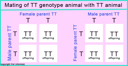
In this diagram, 100% of the offspring will be born TT. They can only inherit the normal T gene from each parent. You can not get cryptorchid-carrier (Tt) or cryptorchidism-affected (tt) offspring from the mating of two normal, non-carrier animals.
Mating a non-cryptorchid, non-cryptorchid-carrier animal with a cryptorchid male (tt) or tt-female animal:
This is the block or square genetics diagram that would be drawn if you mated a male or female TT animal with a male or female tt animal.

In this diagram, 100% of the offspring will be born Tt. They can only inherit a normal T gene from the normal TT parent and they can only inherit an abnormal t gene from the affected tt parent. All of these offspring animals will be
carriers of the defective cryptorchid gene (i.e none of them will be desirable breeding animals), but none of them will outwardly display the cryptorchid trait because each has a dominant normal T gene that will induce normal testicular descent.
Mating a non-cryptorchid, non-cryptorchid-carrier animal with a non-cryptorchid, cryptorchid-carrier animal:
This is the block or square genetics diagram that would be drawn if you mated a male or female TT animal with a male or female Tt animal.
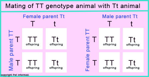
Using this diagram, it is predicted that approximately 50% of the offspring born of this union will be born TT. They can only inherit a normal T gene from the normal TT parent and they have a 50:50 chance of inheriting the normal T gene from the Tt carrier parent. Likewise, approximately 50% of the offspring will be born Tt and become non-cryptorchid carriers of the cryptorchid gene. These animals can only inherit a normal T gene from the normal parent and they have a 50:50 chance of inheriting the abnormal t gene from the Tt carrier parent. Those offspring animals born TT will not have a copy of the defective cryptorchid gene to pass on their own offspring. In this way, by breeding only these TT animals, the cryptorchidism trait can be theoretically eliminated from the breed lines. Those animals born Tt will be
carriers of the defective cryptorchid gene (able to pass it on to future generations), but none of them will outwardly display the cryptorchidism trait because each has a dominant normal T gene that will induce normal testicular descent.
Mating a non-cryptorchid, cryptorchid-carrier animal (Tt) with a non-cryptorchid, cryptorchid-carrier (Tt) animal:
This is the block or square genetics diagram that would be drawn if you mated a male or female Tt animal with a male or female Tt animal.

Using this diagram, it is predicted that approximately 25% of the offspring born of this union will be born TT. They have a 50:50 chance of inheriting a normal T gene from the first carrier parent and they have a 50:50 chance of inheriting a normal T gene from the other carrier parent. Approximately 50% of the offspring will be born Tt and become non-cryptorchid carriers of the cryptorchid gene just like their parents. Finally, approximately 25% of the offspring born of this union will be born tt. They have a 50:50 chance of inheriting the abnormal t gene from the first carrier parent and they have a 50:50 chance of inheriting an abnormal t gene from the other carrier parent. Those 25% of animals born TT will not have a copy of the defective cryptorchidism gene to pass on their own offspring. Those animals born Tt will be carriers of the cryptorchid gene (i.e they will not be desirable breeding animals), but none of them will outwardly display the cryptorchidism trait because each has a dominant normal T gene. The final 25% of animals born tt will outwardly display the cryptorchidism trait if they are males and become dangerous silent carriers of the double-abnormal-gene if they are female (females with the double cryptorchid gene are very dangerous in a breeding population because they pass on the defective gene to 100% of their offspring, but show no outward signs of the condition themselves since they have no testicles). tt animals should not be bred from because they will pass on the defective gene to 100% of their offspring.
Mating a non-cryptorchid, cryptorchid-carrier animal with a cryptorchid male (tt) or tt-female animal:
This is the block or square genetics diagram that would be drawn if you mated a male or female Tt animal with a male or female tt animal (if the tt animal was a male it would be expected to be a clinically cryptorchid animal).
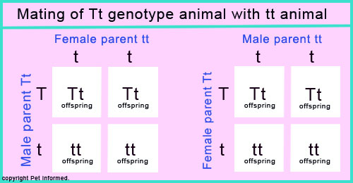
Using this diagram, it is predicted that none of the offspring born of this union will be born with a normal TT genotype. Approximately 50% of the offspring will be born Tt and become non-cryptorchid carriers of the cryptorchid gene. These animals have a 100% chance of inheriting the abnormal t gene from the first tt clinically affected parent and they have a 50:50 chance of inheriting the normal T gene from the other Tt carrier parent. Approximately 50% of the offspring will be born tt. These animals have a 100% chance of inheriting the abnormal t gene from the first tt clinically affected parent and they have a 50:50 chance of inheriting the abnormal t gene from the other Tt carrier parent. Those animals born Tt will be carriers of the cryptorchid gene, but none of them will outwardly display the cryptorchidism trait because each has a dominant normal T gene. The 50% of animals born tt will outwardly display the cryptorchidism trait if they are males and become dangerous silent carriers of the double-abnormal-gene if they are female (females with the double cryptorchidism gene are very dangerous in a breeding population because they pass on the defective gene to 100% of their offspring, but show no outward signs of the condition themselves since they have no testicles). tt animals should not be bred from because they will pass on the defective gene to 100% of their offspring.
Mating a cryptorchid (tt) male with a tt female:
This is the block or square genetics diagram that would be drawn if you mated a male tt animal with a female tt animal. The tt male animal would be expected to be a clinically cryptorchid animal. The female would show no outward signs of being tt-affected.
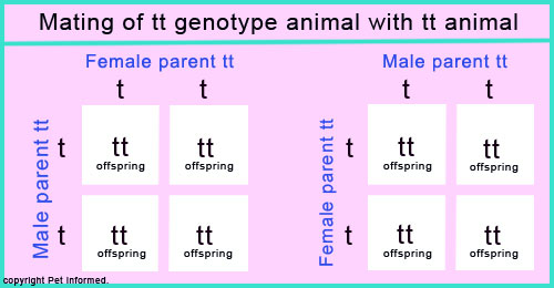
In this diagram, 100% of the offspring will be born tt. They can only inherit the defective t gene
from each parent. You can not get normal TT animals or unaffected, cryptorchid-carrier (Tt) offspring from the mating of two cryptorchid tt animals.
10d) Should I breed from a cryptorchid animal at all?
After reading section 10c on the inheritance of cryptorchidism, it should now be clear to you why the breeding of cryptorchid animals is highly undesirable. Because the cryptorchid condition is a
recessive condition, males that actually display the cryptorchid trait must have two copies of the defective cryptorchid gene (i.e. they must be tt)
in their DNA. The implication of this is that, should these cryptorchidism affected animals be bred from, they will have a 100% chance of passing the defective trait (t) onto 100% of their offspring. All of the offspring produced by mating that cryptorchid animal will be either born affected with cryptorchidism or be carriers of the gene. Consequently, none of the offspring will be considered to be valuable breeding animals themselves.
10e) Should I breed from the parents of a cryptorchid animal?
After reading section 10c on the inheritance of cryptorchidism, it should now be clear to you why the breeding of cryptorchid animals is highly undesirable. What should also be evident is that it requires the mating of two parents, both of whom either carry (Tt) or are affected with (tt) the defective cryptorchid gene, in order for cryptorchidism-affected (tt) male offspring to be produced. The birth of a cryptorchid puppy or puppies should
be a flag to the breeder that
both of the parents are carriers of the defective gene. In order to eliminate the cryptorchidism trait from the breeding population, the parent animals that produced the cryptorchid offspring should no longer be bred from.
10f) Should I breed from the siblings of a cryptorchid animal?
After reading sections 10c and 10e on the inheritance of cryptorchidism, it should now be clear to you that, in order for a cryptorchid animal to be born into a litter, both parents of that affected animal need to be carriers of the defective gene (t). This fact has important breeding implications for the rest of the litter (any other offspring born out of that same t-carrier-animal union that are direct siblings to the cryptorchidism-afflicted male baby). Because the parents are both carriers of the defective gene (either Tt or tt), there is every chance that most of the male and female siblings of the cryptorchid offspring will have inherited and, therefore, be carriers of, the defective (t) gene. The best that can be expected statistically is that a mere 25% of the litter will not carry the defective gene at all and this would require a Tt/Tt parental mating. A Tt/tt mating will result in
all offspring carrying at least one copy of the defective recessive t gene and a tt/tt parent mating will result in
all offspring carrying two copies of the t gene
and being affected with the cryptorchid condition if they are male. Consequently, in order to eliminate the cryptorchidism trait from the breeding population, the siblings of the cryptorchid animal should not be bred from. This includes older siblings born from the previous matings of those two carrier parents.
10g) Is cryptorchidism more common in certain breeds?
The breed of dog or cat may well increase or decrease a veterinarian's suspicions about whether or not it could be cryptorchid. Certain breeds (particularly pure breeds) of dog and cat have a higher incidence of cryptorchidism, whereas certain other breeds have a low incidence of the condition. Dog breeds at high risk of cryptorchidism tend to be small breeds including: Toy and Miniature Poodles, Pomeranians, Chihuahuas, Malteses, Cairn and Yorkshire Terriers, Dachshunds, Schnauzers, shelties (Shetland Sheepdogs), Pekingeses and Bulldogs. Larger breeds, in particular the Boxer and Old English Sheepdog, may also be affected. In the cat world, the Persian is most commonly implicated, however, we have seen a lot of domestic shorthair and longhair cats (moggies) with the condition in our clinic. Large dog breeds seem to have a greatly reduced risk of cryptorchidism. Those at low risk include the Saint Bernard, Great Dane and various field and hunting dogs like the Golden Retriever, Labrador, Beagle and English Setter. Mixed breed (mongrel) dogs are thought to have a lower incidence than pure breed dogs.
10h) Are there drugs or medications or surgical procedures available to make the retained testicle descend (i.e. cryptorchidism repair) and what are the ethical and medical implications of this?
Despite the fact that cryptorchidism is an undesirable hereditary trait, best eliminated through selective castration and non-breeding, there are still many breeders and show-types who refuse to discard a cryptorchid show dog or stud dog and who want their veterinarian to prescribe treatments to descend their animal's testicle or testicles so that they can hide the cryptorchidism fault from potential stud clients, show-judges and dog buyers. Options that have been trialled, with variable success, include both medical options (hormones, drugs) and surgical options.
Medical options:
Most veterinary texts that discuss the matter of drug or hormone-assisted testicular descension suggest the attempt to be, as a general rule, unrewarding. Most veterinary texts say that there is no highly-effective medical treatment available for treating cryptorchidism.
The only report of successful medicine-based induction of testicular descension that I have located comes from the Feldman and Nelson book of Canine and Feline Endocrinology and Reproduction (reference number - 2). This reference indicates that some success in inducing canine (feline?) testicular descension might be achieved by administering either hCG (human chorionic gonadotrophin) or GnRH (gonadotrophin releasing hormone), both of which act on descended or undescended testicular Leydig Cells (Interstitial Cells) to induce them to produce increased quantities of testosterone. The reason these treatments are thought to have some merit is because normal testicular descent (gubernaculum regression) is thought to be mediated by blood testosterone levels. Because cryptorchid testicles usually secrete slightly less testosterone than normally-descended testicles, it is thought that some benefit might be gained by hormonally inducing increased amounts of testosterone to be released.
GnRH: Two doses of GnRH are usually administered at an interval of 1 week apart.
hCG: Four doses of hCG are usually administered over a 2 week period.
There does not seem to be any benefit gained by giving artificial testosterone itself, which is why the GnRH or hCG is used instead.
According to the reference (reference 2), these drugs have only been found to be somewhat successful in the induction of canine testicular descension if they are given to puppies
under 16 weeks of age. Results are very variable and higher success rates seem to be found in dogs whose undescended testicle or testicles are located outside of the abdominal cavity already. Whether the resultant successful testicular descension that occurs is a result of the drugs themselves or a result of the fact that these testicles might have descended anyway is unknown.
Surgical options:
The surgical relocation of an abdominally, inguinally or prescrotally located retained testicle into the scrotal sac is considered to be both a highly-complex and unethical procedure. It is, however, a procedure that is offered to dog breeders and showers by some veterinarians. The surgical procedure is not without potential complications, including the death of the translocated testicle through damage or occlusion of the testicular blood supplies, and there is always the likelihood that the relocated testicle/s will remain infertile, having been previously located at body temperatures warm enough to cause permanent damage to the spermatogenic cells of the testis.
The artificial induction of testicular descent, whether this be through medical or surgical processes, must always be considered questionable from an ethical viewpoint. The condition is a hereditary defect with medical implications for the affected animal and any of its affected offspring. The deliberate breeding of any animal with such hereditary defects should be frowned upon as unethical because of the detriment to the breed's quality overall and because of the suffering and pain (cancer, testicular torsion etc.) that might be experienced by afflicted dogs further down the line.
The practise of inducing testicular descent artificially should also be considered unethical from a business viewpoint too. The whole reason why breeders and showers of dogs and cats seek to get their animal's testicles corrected is to deceive show judges, prospective breeder clients and prospective animal buyers into thinking that their animal is genitally and genetically sound. This results in more ribbons at the show, more stud-dog sire demand and more pups sold to the deceived buyer, but in doing so, it essentially robs the ethical showers of their deserved success and defrauds the owners of breeding bitches and queens who subsequently have their females mated to that prize-winning, but defective stud.
11. Is it possible to place prosthetic testicles (testicle implants) in a cryptorchid animal?
It is possible for veterinary surgeons to place prosthetic testicles into the scrotal sacs of cryptorchid or freshly-neutered animals (dogs and cats) in order to produce the appearance of a normally-descended entire male.
Whether this procedure should be considered ethical or not really depends on the purpose and reasoning behind their use. Placing prosthetic testicles into the scrotal sac of a recently castrated male so that he looks more male to the owner is probably a good thing if it will encourage owners who would not normally have gotten their male pets desexed to do so. After all, there is a big problem with pet overpopulation and animal dumping these days and any product that can help to reduce the incidence of pet breeding and overpopulation is probably a good thing. On the other hand, placing prosthetic testicles into the scrotal sacs of defective, undescended (cryptorchid) males in order to fool show judges, prospective animal buyers and prospective breeding clients into thinking that those males are genetically and genitally sound is unethical. By all means, place prosthetic testicles into cryptorchid dogs and cats if their 'looking normal' pleases you; just make sure that you get those animals castrated at the same time so that they can not breed and make sure that you declare those animals' cryptorchid status to anyone with a vested interest in this matter (e.g. judges at dog shows).
People often ask online whether prosthetic testicle implants are safe. Having not had much experience with prosthetic testicle implants in my years of practice, I can not say for sure. I have to admit, I have never had an Australian owner ask me about testicular implants in a dog or cat, however, this may be mostly due to the fact that I have predominantly worked in emergency medicine, shelter and specialist practices and less-so in high-end general practices where such requests are more likely to be made. According to an Australian breeder friend I know, artificial testicular implants seem to be quite commonly used in Australian show circles and, as far as she knows, no-one seems to be having any problems with them. According to the company (CTI Corporation) that produces the FDA-approved, patented testicular prostheses (called Neuticles), complications with the testicular implants have been very low due to the inert nature of the substances (polypropylene or silicone) that they are made of. After all, similar compounds are used every day in human plastic surgery procedures. What's more, the fact that the manufacturer has backed itself with a multi-million dollar product liability guarantee (i.e. it will compensate owners whose implants do go wrong) suggests that the company is pretty confident of no complications occurring with its product.
For more info on prosthetic testicle implants, go to the "Neuticles" website, run and owned by the CTI Corporation:
http://www.neuticles.com/index.php
Important disclaimer: Please note that this author and website is in
no way affiliated with CTI Corporation: the company that produces testicular implants for pets. Pet Informed's reasons for mentioning the Neuticles website is so that readers interested in reading about or obtaining testicular prosthetics for their pets will know where to turn because, as far as I can tell, CTI Corporation is the only company that has the worldwide patent to produce such products. Pet Informed receives no commercial or reputational benefit from CTI for mentioning the product and can not make any guarantees or claims, either positive or negative, about this company's products, customer service or business practices. Choosing to access testicular implants from any company on the web is your decision alone and Pet Informed can not and will not take any responsibility for any death, damage, injury or loss of reputation and business that occurs should you choose to take this path with your pet. Do your homework and research all such products carefully before spending your money and risking your pet's health on any such products.
To go from this page to the Pet Informed Home Page, click here.
References and Suggested Readings:
1) Clinical and Diagnostic Evaluation of the Male Reproductive Tract. In Feldman EC and Nelson RW: Canine and Feline Endocrinology and Reproduction, 2nd ed. Sydney, 1996, WB Saunders Company.
2) Disorders of the Testes and Epididymides. In Feldman EC and Nelson RW: Canine and Feline Endocrinology and Reproduction, 2nd ed. Sydney, 1996, WB Saunders Company.
3) Feline Reproduction. In Feldman EC and Nelson RW: Canine and Feline Endocrinology and Reproduction, 2nd ed. Sydney, 1996, WB Saunders Company.
4) Reproductive and Hormonal Functions of the Male (and the Pineal Gland). In Guyton AC, Hall JE, editors: Textbook of Medical Physiology, 9th ed. Sydney, 1996, WB Saunders Company.
5) The Digestive Apparatus. In Dyce KM, Sack WO, Wensing CJG editors: Textbook of Veterinary
Anatomy, 2nd ed. Sydney, 1996, WB Saunders Company.
6) The Urogenital Apparatus. In Dyce KM, Sack WO, Wensing CJG editors: Textbook of Veterinary
Anatomy, 2nd ed. Sydney, 1996, WB Saunders Company.
7) The Pelvis and Reproductive Organs of the Carnivores. In Dyce KM, Sack WO, Wensing CJG editors: Textbook of Veterinary
Anatomy, 2nd ed. Sydney, 1996, WB Saunders Company.
8) Surgery of the Reproductive and Genital Systems. In Fossum TW, et al, editors: Small Animal
Surgery. Sydney, 1997, Mosby.
9) Appendix 2. Congenital Defects of the Cat. In Ettinger SJ, Feldman EC, editors: Textbook of Veterinary
Internal Medicine, Sydney, 2000, WB Saunders Company.
10) Appendix 3. Congenital Defects of the Dog. In Ettinger SJ, Feldman EC, editors: Textbook of Veterinary
Internal Medicine, Sydney, 2000, WB Saunders Company.
11) Memon MA, Mickelsen WD, Inherited and Congenital Disorders of the Male and Female Reproductive Systems. In Ettinger SJ, Feldman EC, editors: Textbook of Veterinary
Internal Medicine, Sydney, 2000, WB Saunders Company.
12) Verstegen J, Feline Reproduction. In Ettinger SJ, Feldman EC, editors: Textbook of Veterinary
Internal Medicine, Sydney, 2000, WB Saunders Company.
13) Disorders of the Penis, Prepuce and Testes. In Nelson RW, Couto CG, editors: Small Animal
Internal Medicine, Sydney, 1998, Mosby.
14) Canine Reproduction. In Daris W, editor: Compendium of Animal
Reproduction, 5th ed. 1998, Intervet.
15) Feline Reproduction. In Daris W, editor: Compendium of Animal
Reproduction, 5th ed. 1998, Intervet.
16) Hayes HM et al, Canine Cryptorchidism and Subsequent Testicular Neoplasia: Case Control Study with
Epidemiological Update. In Teratology 32:51, 1985.
Pet Informed is not in any way affiliated with any of the companies whose products appear in images or information contained within this article. The images, taken by Pet Informed, are only used in order to illustrate certain points being made in the article.
Copyright September 14, 2008, www.pet-informed-veterinary-advice-online.com.
All rights reserved, protected under Australian copyright. No images or graphics on this Pet Informed website may be used without written permission of their owner, Dr. O'Meara.
Neuticles is a registered trademark of CTI Corporation.
Please note: the cryptorchidism information provided on this page contains general recommendations and medical advice only. The information provided is based on published information; relevant veterinary literature and publications and my own experience as a practicing veterinarian. The advice given is appropriate to the vast majority of pet owners, however, owners with cryptorchid pets should take it upon themselves to ask their own veterinarian for further advice. Owners with specific circumstances (breeding dogs, showing dogs, stud dogs, breeding businesses, those seeking prosthetic implants etc.) should ask their vet what the safest and most effective protocol is for their situation.
Any dose rates mentioned on these pages should be confirmed by a vet. Dosing rates for common drugs are being changed and updated all the time (e.g. as new research comes in and as drug formulations change) and information here may not remain current for long. What's more, although we try very hard to maintain the accuracy of our information, typos and oversights do occur. Please check with your vet before dosing any pet any medication or drug.
Common misspellings: criptorchidism, criptorchid, cryptorkidism, cryptorkid, criptorkidism, criptorkid,
cryptorkydism,cryptorkyd, criptorkydism, criptorkyd, cryptorchydism, cryptorchyd, criptorchydism, criptorchyd,
testical, testecal, testacal, testucal, testecle, testacle, testucle.
Alternative, slang synonyms for testicles: ball, balls, nut, nuts, goolies.